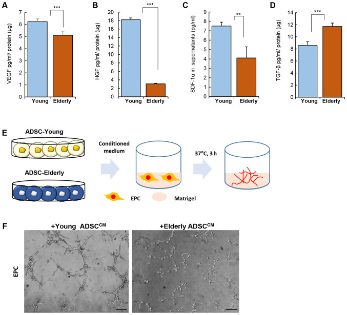
Figure 2.
Effect of age on the paracrine potential of ADSCs. ADSCs were cultured and the culture medium was collected for the quantification of cytokines. Quantification for (A) VEGF, (B) HGF, (C) SDF-1α and (D) TGF-β was performed using ELISA. (E) Experimental scheme for Matrigel assay with EPC. Conditioned medium of ADSC was treated with EPCs on Matrigel. (F) Representative images for tube formation. Data are presented as the mean ± SD of at least three replicates for each group. Scale bar, 100 µm. **P<0.01 and ***P<0.001. ADSCs, adipose-derived stem cells; HGF, hepatocyte growth factor; SDF-1α, stromal cell-derived factor-1α; TGF-β, transforming growth factor-β; VEGF, vascular endothelial growth factor; EPC, endothelial progenitor cell.

