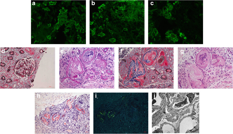
Fig. 1.
Renal biopsy findings of case 1. a showed IgA linear staining along glomerular capillary wall and tubular basement membrane (× 200). b showed λ linear staining along glomerular capillary wall and tubular basement membrane (× 200). c showed λ was strong positive on the protein casts (× 200). d showed minimal mesangial proliferation of the glomeruli (PASM+Masson, × 400). e showed PAS negative protein casts in the tubular lumen (PAS, × 400). f showed fibrillary structure in the peripheral of the protein casts (PASM+Masson, × 400). g showed mononuclear cells in the center of the protein casts (H&E, × 400). h showed the protein casts was Congo red positive (Congo red staining, × 200). i showed the protein revealed apple-green birefringence with polarized microscopy (Congo red staining, × 200). j showed normal glomerular without fine granular deposits along the capillary wall (× 6000)

