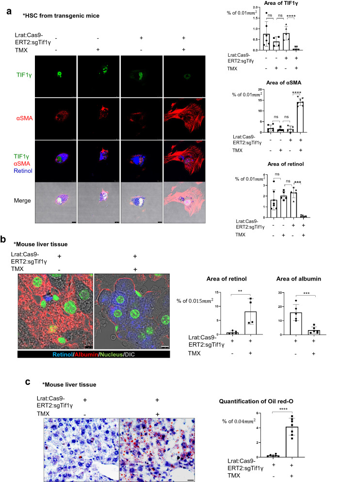
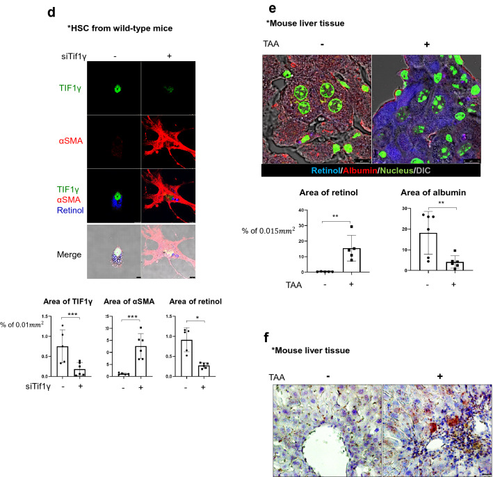
Fig. 1.
The fibrotic liver undergoes a loss of retinol from HSCs and an accumulation of triglyceride in hepatocytes. a Detection of retinol, TIF1γ, and αSMA in primary hepatic stellate cells from Lrat:Cas9-ERT2: sgTif1γ mice treated with or without 10 nM TMX in DMSO for 3 days. The control group was treated with DMSO only. TIF1γ is shown in green, αSMA is shown in red, and retinol is shown in blue. TIF1γ or αSMA-positive area % per 0.01 mm2 were quantified in five images each group using Image J program. Reproducible result from two independent experiments was shown. ***P < 0.001, ****P < 0.0001, ns; non-significant. Scale bar: 10 μm. b Detection of retinol (blue) and albumin (red) in the liver tissue of normal and Lrat:Cas9-ERT2: sgTif1γ mice treated with TMX in DMSO. The nucleus was stained with SYTOX green. DMSO only in TG, n = 4; TMX in TG, n = 4, complied from two experiments. Retinol or albumin-positive area % per 0.015 mm2 were quantified using Image J program. **P < 0.01, ***P < 0.001. Scale bar: 5 μm. c Oil red-O staining of triglyceride distribution in the liver of Lrat:Cas9-ERT2: sgTif1γ mice. The nuclei were stained with hematoxylin. DMSO only in TG, n = 4; TMX in TG, n = 4, complied from two experiments. Oil-red-O-positive area % per 0.04 mm2 were quantified using Image J program. ****P < 0.0001. Scale bar: 2 μm. d Detection of retinol, TIF1γ, and αSMA in primary hepatic stellate cells from normal mice treated with or without siTif1γ. TIF1γ is shown in green, αSMA is shown in red, and retinol is shown in blue. TIF1γ or αSMA-positive area % per 0.01 mm2 were quantified using Image J program. Reproducible result from two independent experiments was shown. *P < 0.05, ***P < 0.001, Scale bar: 10 μm. e Detection of retinol (blue) and albumin (red) in the liver tissue of normal and treated with TAA (n = 4 each group, two experiments). The nucleus was stained with SYTOX green. Retinol or albumin-positive area % per 0.015 mm2 were quantified using Image J program. **P < 0.01, Scale bar: 5 μm. f Oil red-O staining of triglyceride distribution in the normal and TAA-induced mouse liver (n = 4 each group, two experiments). Scale bar: 2 μm


