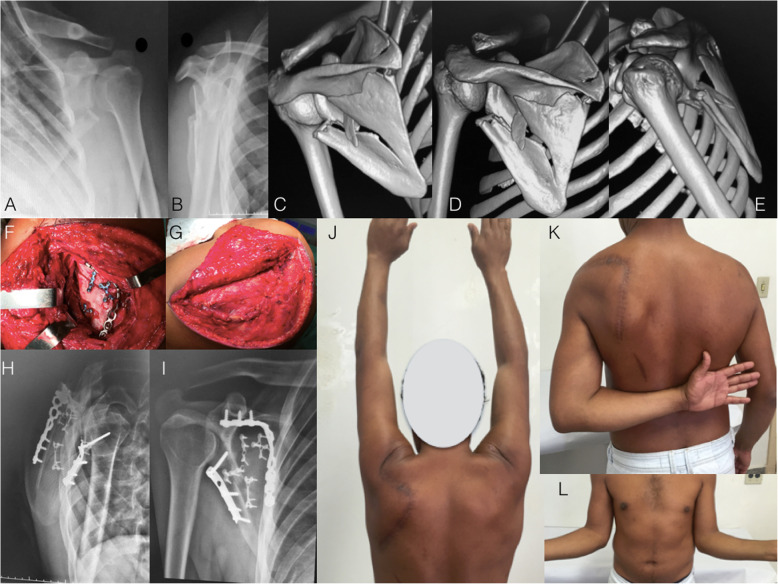
Fig. 1.
a and b: Radiographs of the left shoulder of a 34-year-old male patient who suffered a motorcycle accident and presented a severely displaced and comminuted infraspinous fracture of the scapula body. c, d, and e: Computed tomography with three-dimensional reconstruction. Observe the angulation of the scapula body and the degree of glenoid medialization. f: Perioperative photography depicting the classic Judet approach. Observe the extensile detachment of the infraspinatus muscle. g: Perioperative photography showing the closure and infraspinatus muscle. h and i: Radiographs after three months showing the fracture healing after fixation with one-third tubular plate at the lateral pillar and a twisted reconstruction plate at the medial pillar of the scapula. Fragment-specific fixation using 2.0-mm minifragment plates was also performed. j, k, and l: Postoperative photographs showing complete range of motion recovery after three months of surgery

