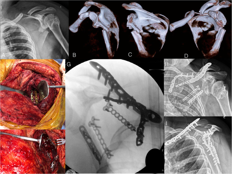
Fig. 2.
a: Radiograph of the right shoulder in anteroposterior view of a 24-year-old male patient who suffered a car accident and presented a severely displaced midshaft clavicle fracture in combination with an infraglenoid fracture of the scapula body. Observe that the patient presented a sequelae of previous proximal humeral and glenoid fractures, with no residual shoulder instability. b, c, and d: 3-D CT reconstruction showing the medialization of the glenoid and the angulation of the scapular body. e and f: Perioperative photographs depicting the modified Judet approach. Observe the fixation of the lateral pillar of the scapula with two plates at the interval between the infraspinatus and teres minor muscles (e). The medial pillar of the scapula was reduced and fixed with a twisted reconstruction locking plate. Observe the minimal detachment of the infraspinatus muscle (f). g: Perioperative fluoroscopy image showing scapula and clavicle fractures reduction and fixation. h and i: Radiographs in anteroposterior and lateral views showing fracture healing after three months

