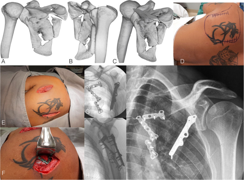
Fig. 3.
a, b, and c: 3-D CT reconstruction showing a comminuted infraglenoid fracture of the scapular body in a 35-year-old male patient. Observe the angulation of the inferior part of the scapular body and the medialization of the glenoid. d: Preoperative photography depicting the landmarks for minimally invasive approach. e and f: Perioperative photographs showing the lateral (between infraspinatus and teres minor muscles) and medial approaches (partial detachment of the infraspinatus). g and h: Postoperative fluoroscopy images in anteroposterior and lateral views showing fracture reduction and fixation using 2.7 minifragment plates (medial pillar) and the unconventional use of a 2.7 fibular plate (lateral pillar)

