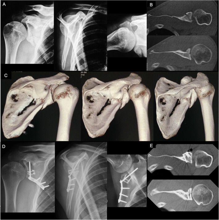
Fig. 6.
a: Preoperative true AP, lateral scapular, and axillary radiographic views of the right shoulder of a 25-years-old male patient, showing a displaced inferior glenoid fragment extending to the lateral pillar of the scapular neck and body. Patient reported on a fall from stairs 48 h before. Also note the small bone fragments in the inferior portion of the capsule (yellow arrowheads); b: Preoperative CT axial cuts of the right shoulder demonstrating the displaced inferior glenoid fracture; c: Preoperative 3-D CT reconstructions showing the displaced inferior glenoid fracture extending to the lateral pillar of the scapular neck and body; d: Postoperative true AP, lateral scapular, and axillary radiographic views of the right shoulder demonstrating the anatomic reduction of the inferior glenoid fracture and buttressing with a one-third tubular plate. Observe the 2.3-mm reduction plate used to maintain the reduction during surgery. Note the long 3.5-mm screw inserted through the plate directed to the coracoid process; e: Postoperative CT axial cuts of the right shoulder demonstrating the anatomic reduction of the inferior glenoid fracture

