Fig. 1.
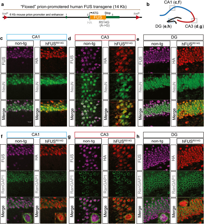
ALS-linked mutant FUS transgene is restricted to neurons and has cytosolic localization. a Schematic of FUS transgene. Murine prion promoter was used to drive the HA-tagged human FUS cDNA expression (R514G). b Schematic representation of different subregions (CA1, CA3 and DG) within hippocampus, and used for panels c–h. Confocal images of endogenous FUS (magenta) and human FUS transgene (red) in the CA1 (c), CA3 (d) and DG (e) of the hippocampus at 3 months of age. Transgenic FUS protein was detected using a HA antibody. Both endogenous FUS and human FUS transgene were present in the neuronal nucleus. Scale bar is 20 μm. Confocal images of the CA1 (f), CA3 (g), and DG (h) regions that were co-labeled with endogenous FUS (magenta) or R514G-FUS transgene (red) and nuclear envelope marker (green) from non-transgenic and R514G mice at 3 months of age. Endogenous FUS (magenta) is restricted to nuclei, whereas R514G-FUS transgene (red) showed both nuclear and cytosolic distribution. The nuclear envelope appeared to be normal across all regions of the hippocampus. Scale bar = 20 μm

