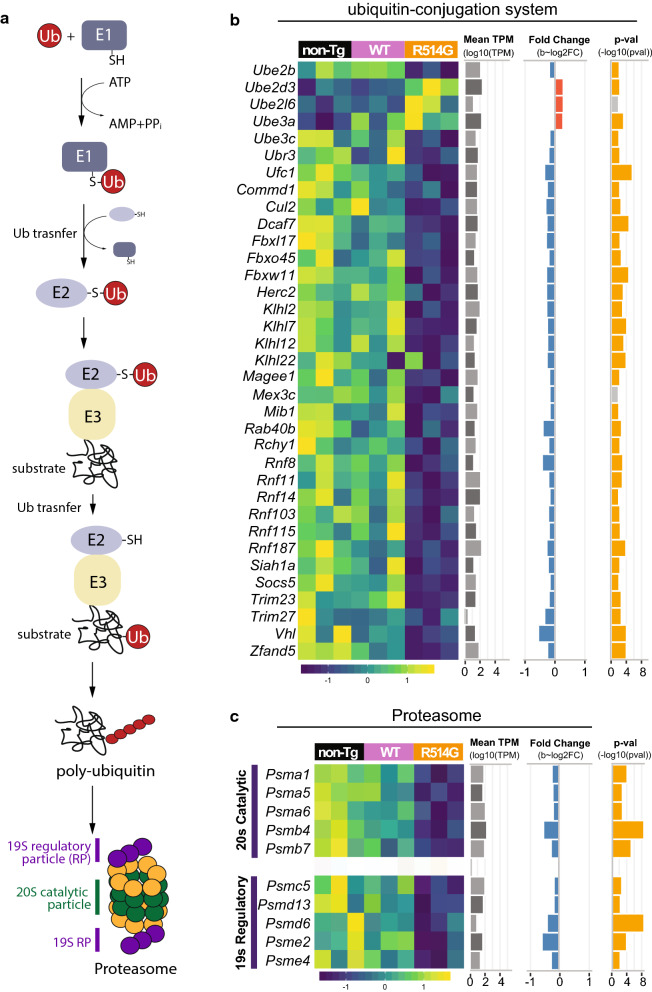
Fig. 8.
Defected ubiquitin–proteasome axis in R514G mice. a Simplified schematic of ubiquitin–proteasome pathway. Ubiquitin is attached to the substrate via E1, E2 and E3. The poly-ubiquitinated proteins were targeted to proteasome for degradation. Proteasome is composed of 20S catalytic particle and 19S regulatory particle (RP). b Heat map of the expression level of the DEGs for ubiquitin–proteasome pathway in non-transgenic, WT-FUS and R514G-FUS mice along with their mean expression level across all samples (log10(TPM)), and p-value (− log10(p-value)) and fold change (log2(fold change)) in R514G-FUS mice as compared to non-transgenic mice. Positive and negative fold changes are colored red and blue respectively and p-values corresponding to significant q-values < 0.1 are colored in yellow

