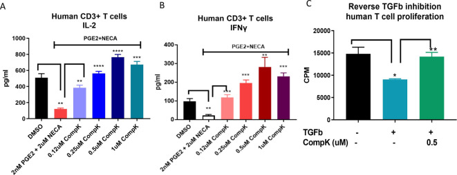
Figure 2.
Improved function of T cells by CompK under immunosuppressive conditions. (A) Recovery of IL-2 release from human primary T cells by CompK in the presence of PGE2 and NECA. Purified human T cells were prereated with PGE2 and NECA for 24 hours in the presence of anti-CD3 and anti-CD28. CompK was then added to the cell culture for an additional 48 hours followed by measurement of IL-2 production. (B) Reinvigoration of IFN-γ production by CompK under suppressive conditions created by PGE2 and NECA. The treatment conditions were the same as IL-2 evaluation. (C) Attenuation of TGF-β inhibition on T-cell proliferation by CompK. Purified human T cells were treated with 3 ng/mL TGF-β for 72 hours in the presence of 0.5 µM CompK. The cells were pulsed with 3H-labeled thymidine 6 hours prior to harvest. All studies were repeated with T cells from multiple donors. The representative graphs are shown here. Student t-test was used for statistical analysis. *P≤0.05, **P<0.01, ***P<0.001, ****P<0.0001. CompK, compound K; DMSO, dimethyl sulfoxide; IFN-γ, interferon gamma; IL, interleukin; NECA, 5′-(N-ethylcarboxamido) adenosine; TGF-β, transforming growth factor beta.

