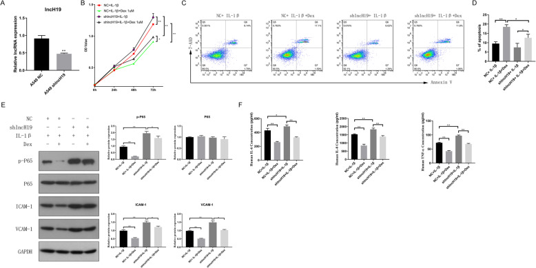
Fig. 4.
When lncH19 was inhibited but cells were treated with IL-1β (10 ng/mL) with or without Dex (1 μM) at the same time, cell proliferation increased, cell apoptosis decreased, and the protein levels of inflammatory genes increased, promoting the phosphorylation of P65, ICAM-1, VCAM-1, and inflammatory cytokines. a. lncH19 expression was reduced, indicating that an lncH19-inhibited cell line was generated. b. Cell proliferation of A549 NC and A549 shlncH19 cells treated with IL-1β with or without Dex. c. Representative cell apoptosis diagram, as measured via flow cytometry. Upper left is the fragment and damaged cells, upper right is the late apoptosis and dead cells, lower left is the normal cells of negative control, and lower right is the early apoptotic cells. The total percent of apoptosis cells was calculated by the sum of cells in the upper right and lower right. d. Percentage of apoptotic cells. e. Western blotting for determining the protein levels of inflammatory genes. f. ELISA for assessing the levels of inflammatory cytokines. Data are presented as mean ± SD. N = 3. *P < 0.05, **P < 0.01. Dex: dexamethasone, IL-1β: interleukin-1β

