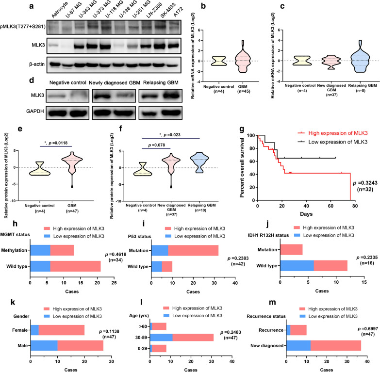
Fig. 1.
Expression pattern of MLK3 in glioblastoma and the correlation between MLK3 expression and patients’ prognosis and other clinical information. a Protein expression and phosphorylation of MLK3 in a set of GBM cell lines. Human astrocytes were used for comparison. Expression of β-actin was served as a loading control. b, c mRNA expression of MLK3 gene in GBM specimens from tumor bank of the Shenzhen Second People’s Hospital. Two grade I gliomas (including 1 angiocentric glioma, 1 pilocytic astrocytoma) and two gliosis were used as the negative control. d Representative immunoblots of MLK3 protein expression in GBM specimens. e, f Overexpression of MLK3 was significant in 47 GBM samples, especially in 10 relapsing GBM samples. P values were determined by Mann–Whitney U test or Kruskal–Wallis one-way ANOVA with Dunn's multiple comparisons test. *: p < 0.05; (G) Kaplan–Meier survival analysis of GBM patients categorized by MLK3 expression and statistical comparisons using Log-rank test. h–l Chi-square test (Fisher exact test) was used to evaluate the correlation between MLK3 protein expression and clinical information (MGMT methylation, p53 mutation, IDH1 mutation, gender, age, and recurrence) of GBM patients

