Fig. 3.
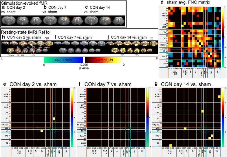
Functional MRI of concussed and sham animals. a–c Post-concussion stimulus-evoked fMRI activity changes of a CON day 2 versus sham, b CON day 7 versus sham, c CON day 14 versus sham. h–j Post-concussion changes in local intrinsic functional connectivity in (H) CON day 2 versus sham, (I) CON day 7 versus sham, and j CON day 14 versus sham. a–c, h–j Statistical map thresholded at p value < 0.05 (two-tailed), unpaired two sample t test, implemented as permutation tested for the General Linear Model, corrected for multiple comparisons with mass-based FSL’s Threshold-free Cluster enhancement (TFCE). Statistical maps were overlaid on the study-specific averaged EPI images. Red anatomical orientation marker L = Left, R = Right. ACA = Anterior Cingulate Area, AM = Amygdala, AUD = Auditory Area, CPu = Caudate Putamen, GP = Globus Pallidus, HP = Hippocampus, INS = Insula, M2 = Secondary Motor Cortex, MB = Midbrain, PRT = Pretectal area, S1 = Primary Somatosensory Cortex, RSN = Retrosplenial Area, TH-pmc = Thalamus-polymodal association cortex, VIS = Visual Area. d Average functional network connectivity (FNC) matrices among Independent Components (ICs) identified by IVA-GL of (a) sham (n = 14). Colour scaled by z test statistics; non-black cells were defined as component–component connectivity deemed statistically significant. One sample t tests, permutation-tested, and FDR-corrected (q value < 0.05, two-tailed). e–g Post-concussion FNC changes of e CON day 2 versus sham, f CON day 7 versus sham, g CON day 14 versus sham. Colour scaled by z test statistics; non-black cells were defined as component–component connectivity deemed statistically significant. Two sample t tests, permutation-tested, and FDR-corrected (q value < 0.1, two-tailed)

