Fig. 2.
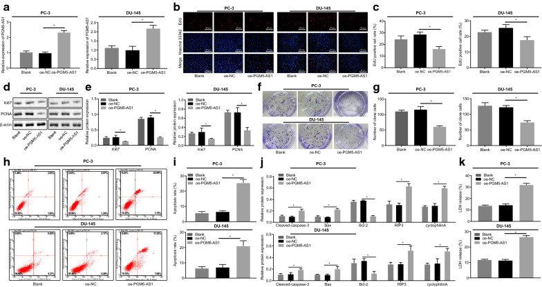
PGM5-AS1 overexpression inhibits the proliferation and colony formation of PC-3 and DU-145 cells and promotes their apoptosis. a The relative expression of PGM5-AS1 in PC-3 and DU-145 cells after transfection through RT-qPCR assay. b The representative images of cell proliferation in transfected PC-3 cells as measured by EdU assay (×200). c The quantitative analysis of cell proliferation in transfected PC-3 cells as measured by EdU assay. d The protein bands of expression of Ki67 and PCNA in transfected PC-3 cells through Western blot analysis. e The quantitative analysis of expression of Ki67 and PCNA in transfected PC-3 cells through Western blot analysis. f The representative images of cell colony formation in transfected PC-3 cells. g The quantitative analysis of cell colony formation in transfected PC-3 cells. h The flow cytometric detection of cell apoptosis in transfected PC-3 cells. i The quantitative analysis of cell apoptosis in transfected PC-3 cells detected by flow cytometric analysis. j The quantitative analysis of expression of cleaved caspase-3, Bax, RIP3, cyclophilinA and Bcl-2 in transfected PC-3 cells through Western blot analysis. k LDH release determined by ELISA. *p < 0.05 vs. the oe-NC group (PCa cells treated with oe-NC). The measurement data were summarized as mean ± standard error and analyzed using one-way ANOVA, followed by Tukey's post hoc test. The experiment was repeated three times independently

