Fig. 6.
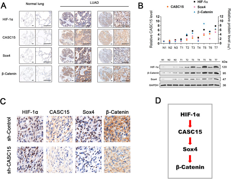
HIF-1α/CASC15/SOX4/β-catenin pathway is activated in a substantial subset of NSCLC patients and A549 xenograft tissues. a Representative ISH staining of CASC15 and IHC staining of HIF-1α, SOX4, and β-catenin in a tissue microarray consisting of 35 matched pairs of NSCLC and adjacent normal lung tissues. Scale bar: 200 μm. b Positive correlation between CASC15 expression and levels of HIF-1α, SOX4, and β-catenin in NSCLC and adjacent normal lung tissues. LncRNA expression was evaluated by qRT-PCR, and protein abundance was evaluated by Western blotting. c Representative ISH staining of CASC15 and IHC staining of HIF-1α, SOX4, and β-catenin in A549-shControl and A549-shCASC15 xenograft tissues. d A schematic model of HIF-1α/CASC15/SOX4/β-catenin signaling pathway activated in NSCLC

