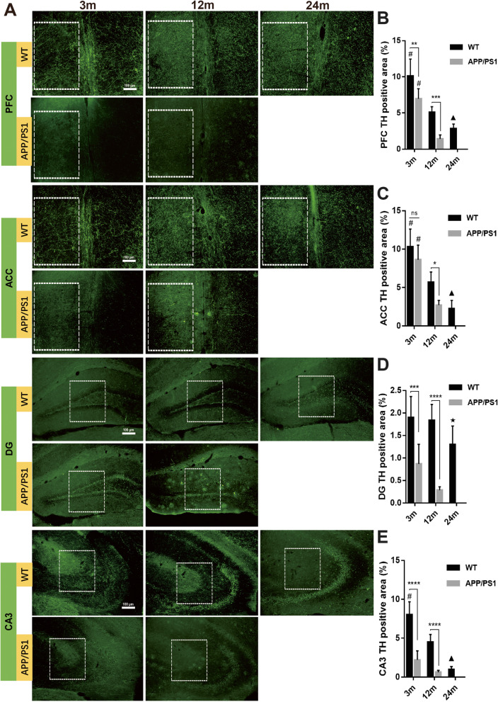
Fig. 6.
Detection of TH+ nerve fibers in the brains of APP/PS1 and WT mice. a Immunofluorescence detection of TH+ nerve fibers in different brain regions showed that in the b medial prefrontal cortex, c anterior cingulate, d DG, and e CA3, TH+ nerve fibers gradually decreased with aging. In 12-month-old APP/PS1 mice, TH+ area was significantly reduced compared with WT mice of the same age and 3-month-old APP/PS1 mice. TH+ area in the brain slices of 3-month-old and 12-month-old APP/PS1 mice was significantly lower than that of WT mice of the same age. In DG, TH+ area of 12-month-old APP/PS1 mice was even significantly lower than that of 24-month-old WT mice. White box indicated region of interests used for the calculation of TH+ area. n = 6. ns, not significant. *P < 0.05, **P < 0.01, ***P < 0.001, ****P < 0.0001, #P < 0.05 compared with other time points in the same group. ▲P < 0.05 compared with 12 m WT mice. ★P < 0.01 compared with 12 m APP/PS1 mice

