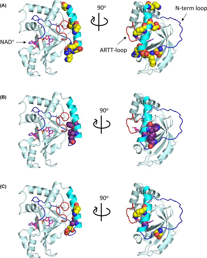
Figure 3. Location of N-terminal motifs in the C3larvinA homology model.
The C3larvinA homology model is shown in cartoon and colored pale cyan. The ARTT-loop is shown in red; the α1-helix is shown in cyan; the unstructured N-terminus is shown in blue. Using the structural alignment tool in PyMOL ver. 1.3, the NAD+ substrate (magenta) was modeled into the active site of C3larvinA using the C3bot1–NAD+ complex (PDB ID: 2C8F) as a template. (A) Residues of the B-motif, D23/K25/D27/R28/K36/E38/K45, are shown using the space-filling model and are colored yellow. (B) Residues of the S-motif, F24/A31/W34, are shown using the space-filling model and are colored purple. (C) The residues of interest, Asp23, Ala31 and Lys36, are shown using the space-filling model and colored based on their respective motifs (D23 and K36 in yellow; A31 in purple).

