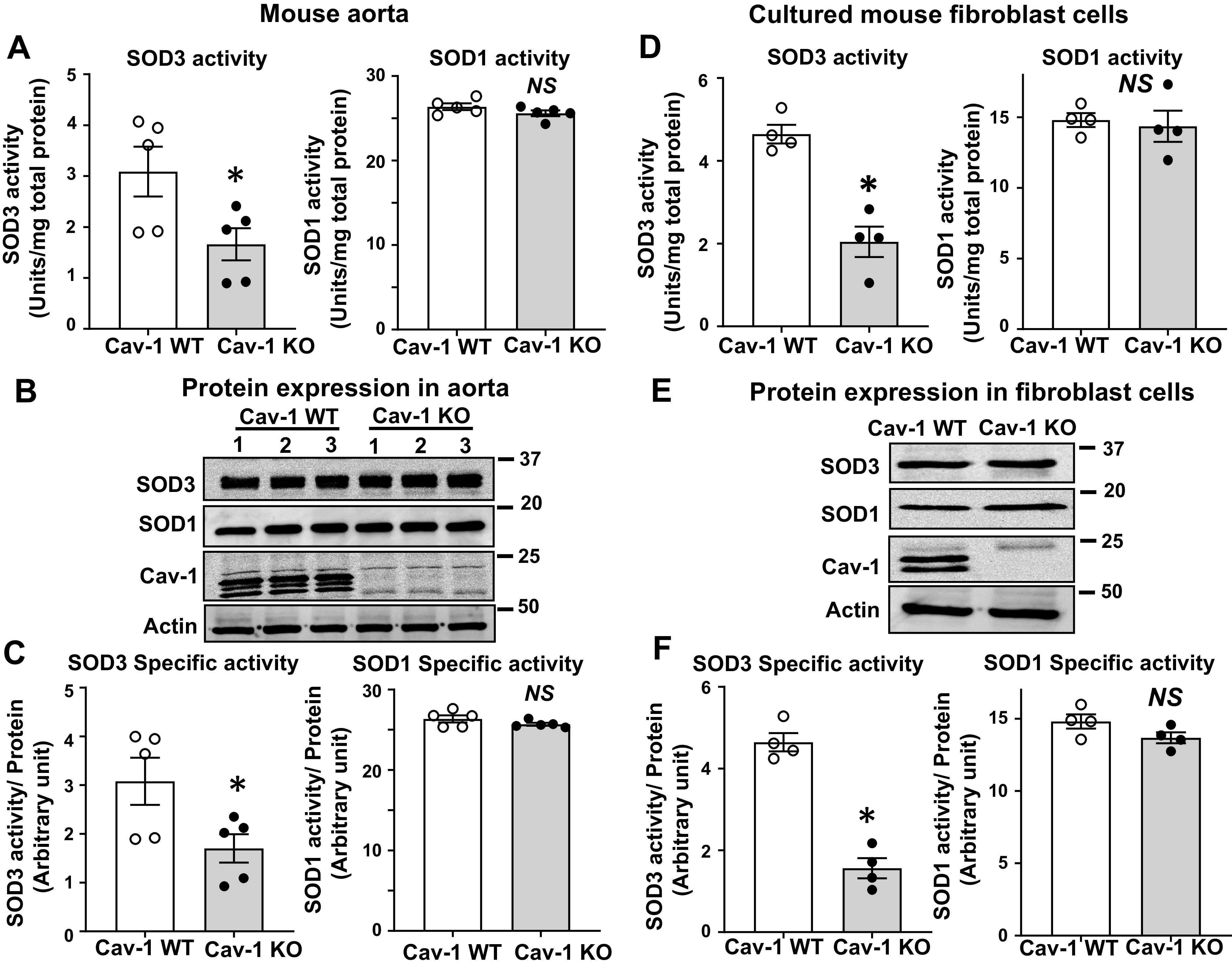
Fig. 1.

Specific activity of superoxide dismutase (SOD3) is decreased in vascular tissue and cultured fibroblast from caveolin-1 knockout (KO) mice (Cav-1−/−). A: activity of SOD3 and SOD1 in aortae from Cav-1 wild-type (WT) and Cav-1−/− mice as measured by inhibition of cytochrome c reduction in the presence of xanthine/xanthine oxidase. Concanavalin A (Con A)-Sepharose chromatography was used to isolate SOD3 from tissue homogenates. B: protein levels of SOD1, SOD3, Cav-1, and actin in aortic tissue. C: the specific activity of SOD1 and SOD3 was determined by the ratio of SOD activity relative to the amount of protein (n = 5). D–F: mouse fibroblast cells were cultured in 1% serum containing DMEM for 72 h. SOD3 secreted into the culture medium was collected and concentrated by concanavalin A-Sepharose chromatography. Activity of SOD3 in concentrated culture medium and SOD1 in cell lysates was assayed (D). Protein levels of SOD1 in cell lysate and SOD3 from conditional medium were determined by Western analysis (E). Specific activity of SOD1 and SOD3 was determined by the relative ratio of SOD activity to the amount of protein (n = 4) (F). Results are presented as means ± SE. *P < 0.05 vs. control. NS, not significant.
