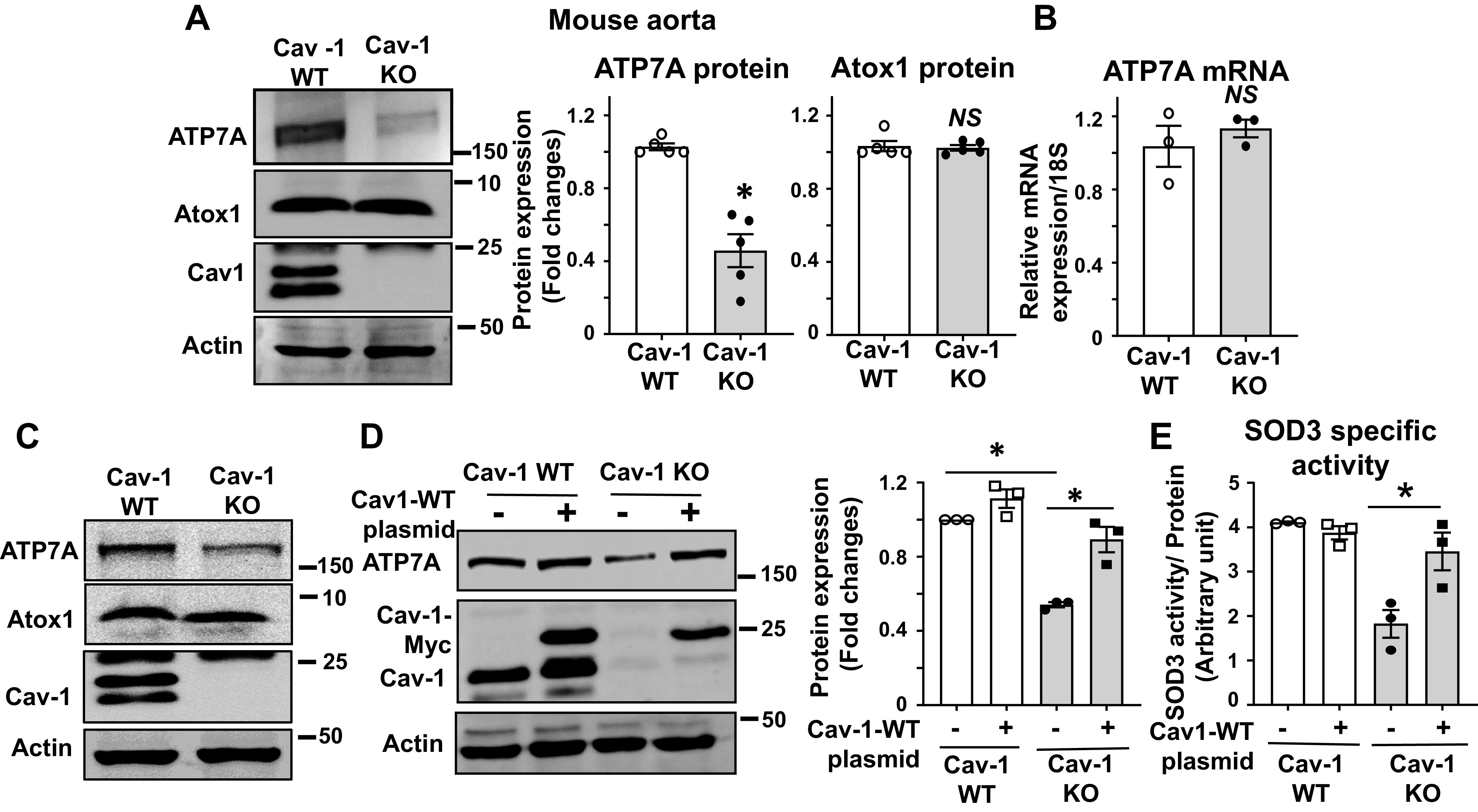
Fig. 4.

Protein expression of the copper transporter ATP7A is decreased in blood vessels from caveolin-1 knockout (KO) mice (Cav-1−/−). A: relative protein expression of ATP7A and Atox1 in aortas from Cav-1 wild-type (WT) and Cav-1−/− mice was determined by Western blotting with antibodies specific to respective proteins. Densitometric analysis is shown (right, n = 5). B: ATP7A mRNA expression in aortas from Cav-1 WT and Cav-1−/− mice was determined by real-time quantitative RT-PCR (n = 3). C: protein expression for ATP7A and Atox1 in mouse fibroblasts isolated from Cav-1 WT and Cav-1−/− mice (n = 3). D: Cav-1 WT and Cav-1−/− mouse fibroblasts mice were transfected with Cav-1 WT plasmid. Lysates were used to measure ATP7A, Cav-1, and actin protein expression. (n = 3). E: superoxide dismutase (SOD3)-specific activity in conditional medium of Cav-1 WT plasmid transfected Cav-1−/− mouse fibroblast cells. Cav-1 WT and Cav-1−/− mouse fibroblast cells were transfected with Cav-1-WT plasmid. The specific activity of SOD3 was determined by the ratio of activity to relative amount of protein in culture conditional medium. Results are presented as means ± SE. *P < 0.05, NS, not significant. ATP7a, Menkes ATPase, copper-transporting P-type ATPase.
