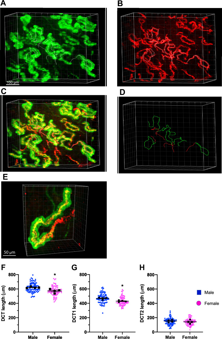
Fig. 3.
Structural sexual dimorphism of the early distal convoluted tubule (DCT1) and late DCT (DCT2). A−C: three-dimensional views of the DCT in common kidney sections colabeled with antibodies to parvalbumin (A; green, DCT1) and Na+-Cl− cotransporter (NCC; B; DCT1 and DCT2) as well as the merged imaged overlay of both green and red (C). D: line tracing of a representative DCT1 (green) and DCT2 (red only) in a section. E: higher-magnification view of a representative single DCT labeled with parvalbumin (green) and NCC (red). F–H: quantification of DCT (F), DCT1 (G), and DCT2 lengths (H) in male and female mice at the basal state. The NCC-positive labeled section of the tubule represents total DCT length; tubules colabeled with NCC and parvalbumin are DCT1 and single labeling with NCC are DCT2. Colored dots are individual measurement of a signal tubule. Black squares are average measurements per kidney. Data are reported as means ± SE; n = 4 kidneys/group and ∼25 DCTs/kidney). Student t tests were performed, and *P < 0.05 was considered statistically significant.

