Figure 1.

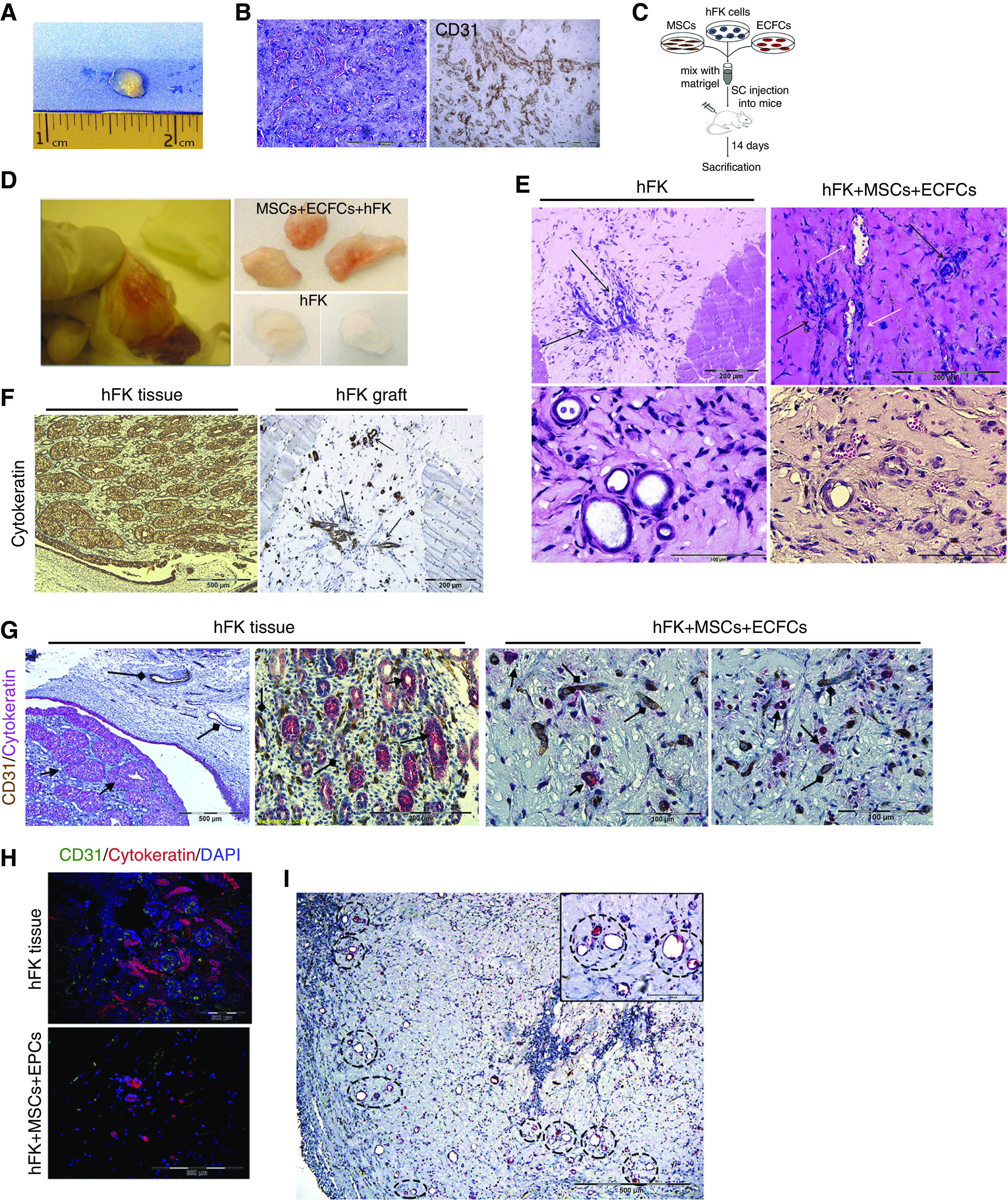
Subcutaneous coadministration of hFK cells, MSCs, and ECFCs results in the formation of renovascular units. (A and B) First, MSCs and ECFCs (106 and 7.5×10⁵, respectively) were mixed with Matrigel and injected into NOD-SCID mice. (A) Seven days later, the grafts were removed for analysis. (B) H&E staining showed patent vascular networks filled with erythrocytes (left). Anti-human CD31 staining confirmed that the vessels’ origin was from the injected MSCs/ECFCs (right). Scale bars, 200 μm. (C) Experiment scheme: to test whether adding hFK cells would allow generation of renal tubules, NOD-SCID mice were injected with either hFK cells alone (106 cells/graft) or with a mix of hFK cells, MSCs, and ECFCs, within Matrigel, and euthanized 14 days later. (D) Macroscopically, mixed grafts exhibited a vascularized appearance (left and top right), compared with hFK only (bottom right). (E) H&E staining showed the presence of tubular structures in both groups (black arrows). However, implants from the mixed group also harbored blood vessels (white arrows). Scale bars: top, 200 μm; bottom, 100 μm. (F) Anti-human cytokeratin was used to track hFK cells in the grafts. hFK tissue served as positive control (left). We detected cytokeratin positive tubular structures (arrows). Scale bars: left, 500 μm; right, 200 μm. (G) Staining of mixed grafts (right) for human CD31 (brown) and cytokeratin (red), demonstrating the presence of both blood vessels (diamond arrows) and tubular structures (arrows). hFK tissue (left) served as a positive control. Scale bars: left, 500 μm; middle left, 200 μm; middle right and right, 100 μm. (H) Immunofluorescence staining of mixed grafts (bottom) for human CD31 (green) and cytokeratin (red), confirming the copresence of human blood vessels with hFK cell–derived tubular structures. hFK tissue (top) served as a positive control. Scale bars, 200 μm. (I) Within the grafts, donor-derived vessels and tubules formed in close proximity (H&E staining with renovascular units marked by circles, inset showing larger magnification of two such units). Scale bar, 500 μm; inset, 100 μm. SC, subcutaneous.
