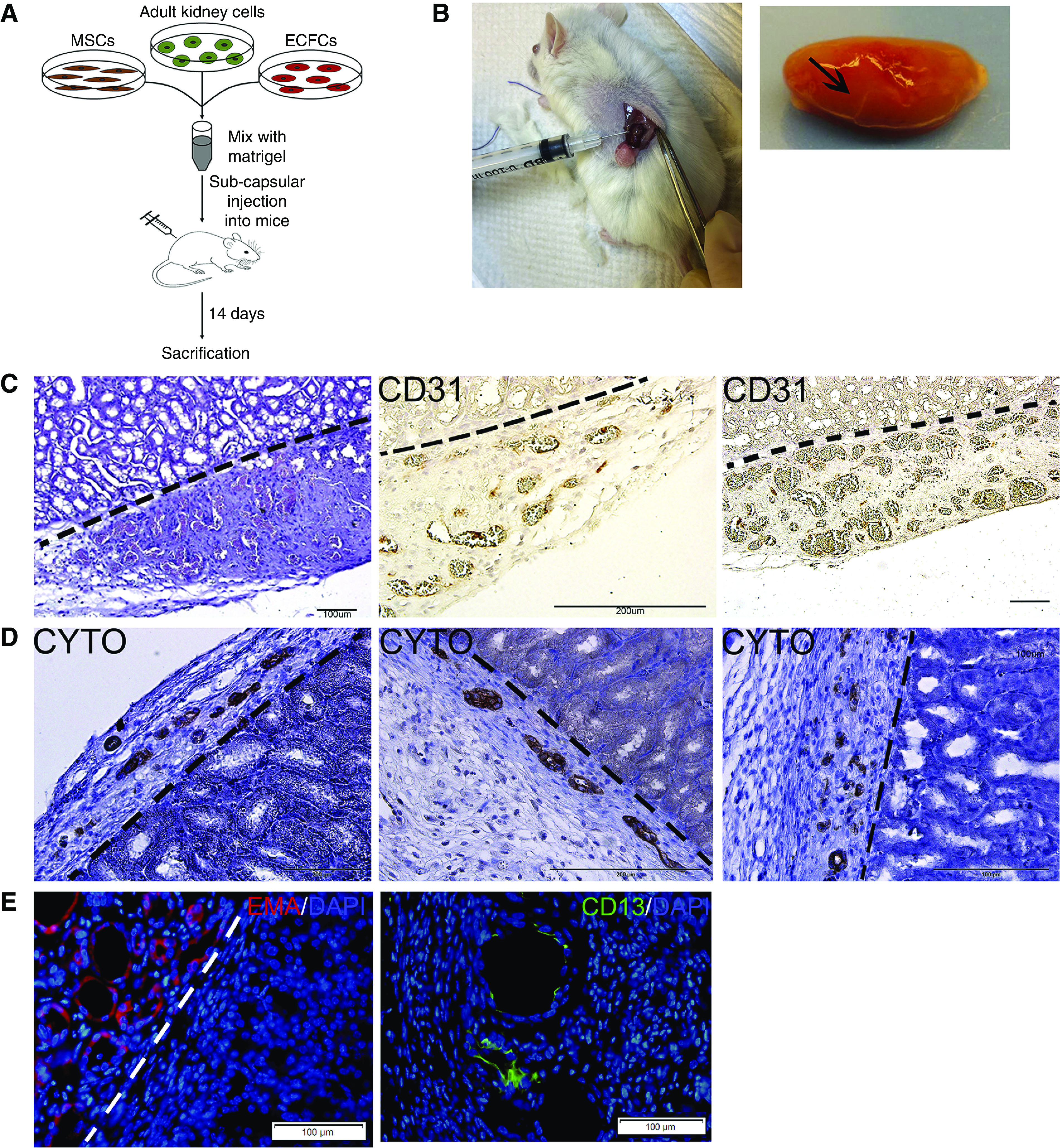
Figure 4.

Subcapsular coadministration of hAK cells, MSCs, and ECFCs results in formation of renovascular units. (A) Experimental scheme. (B) Macroscopic appearance of the grafts in situ (left) and after removal (right, graft marked by arrow on the whole resected kidney). (C) H&E staining (left) and staining for human CD31 (middle and right) demonstrate the formation of erythrocyte-filled, donor-derived vessels. Scale bars: left and right, 100 μm; middle, 200 μm. (D) Staining for cytokeratin demonstrates the formation of tubular epithelial structures. Scale bars: left and middle, 200 μm; right: 100 μm. (E) Immunofluorescence staining for the distal tubule marker EMA (left) and proximal tubule marker CD13 (right), demonstrating the formation of renal tubules of both lineages. Dotted lines mark the border between the graft and mouse kidney tissue.
