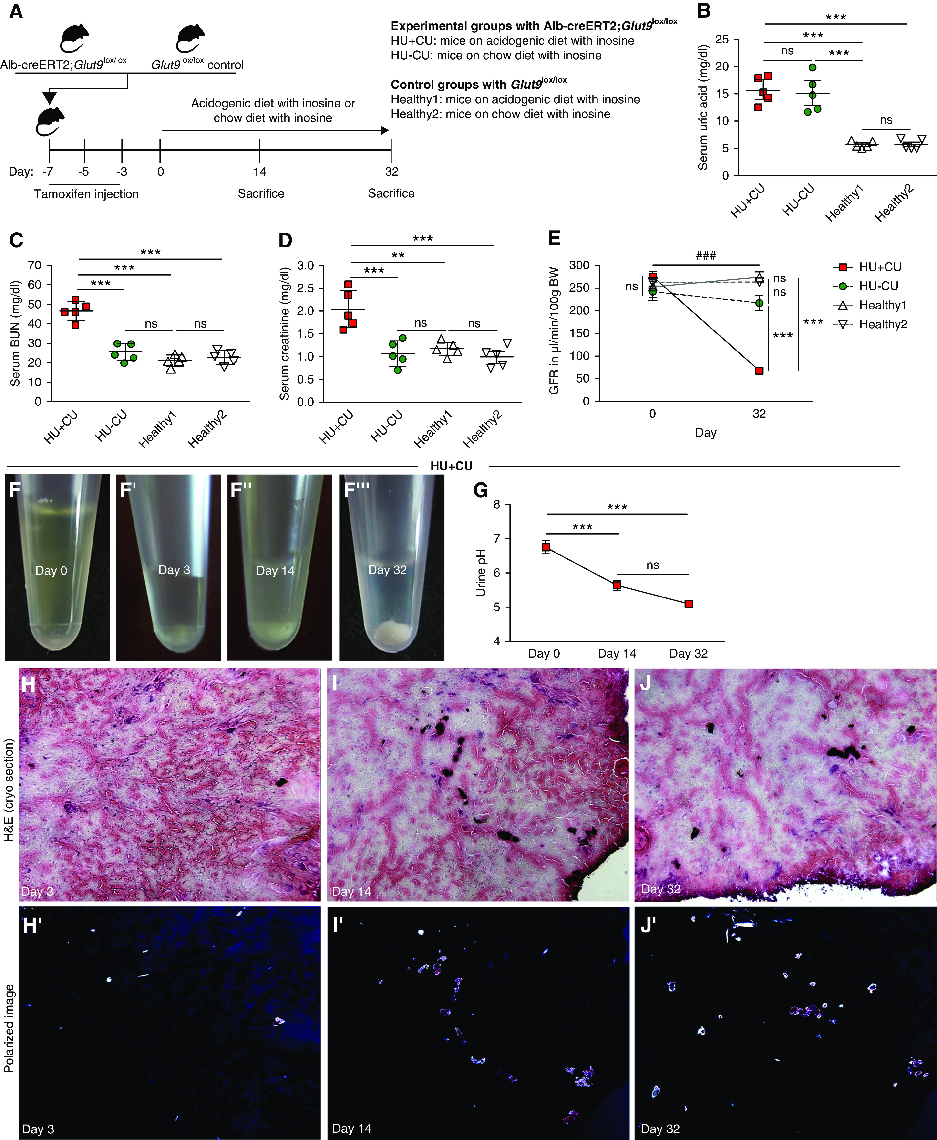
Figure 2.

Renal crystal deposits lead to CKD in mice. (A) Alb-creERT2;Glut9lox/lox and Glut9lox/lox control mice were injected intraperitoneally with tamoxifen. Both groups were fed either an acidogenic diet enriched with inosine or a standard chow diet with inosine up to 32 days. (B–D) Serum UA (B), BUN (C), and creatinine (D) levels of Alb-creERT2;Glut9lox/lox mice with acidogenic diet and inosine (HU+CU) or chow diet with inosine (HU−CU), and Glut9lox/lox mice with acidogenic diet with inosine (healthy2) or chow diet with inosine (healthy1) on day 32 (n=5 mice per group, using one-way ANOVA). (E) GFR of all four groups from day 0 to day 32 (n=5 mice per group, two-way ANOVA). (F) Urine was collected from HU+CU mice that were fed an acidogenic diet with inosine on days 0, 3, 14, and 32. Images illustrate UA crystals in urine on days 3, 14, and 32 (F’–F’’’). (G) Urinary pH was measured from HU+CU mice using a pH meter (n=5 mice per group, one-way ANOVA). (H–J) H&E staining of frozen unfixed kidney sections (H–J) and crystal deposits visualized under a polarizable light microscope from Alb-creERT2;Glut9lox/lox mice with acidogenic diet and inosine (HU+CU) on days 3, 14, and 32 (H’–J’). Data are mean±SD. **P<0.01; *** or ###P<0.001; NS, not significant.
