Fig 1.
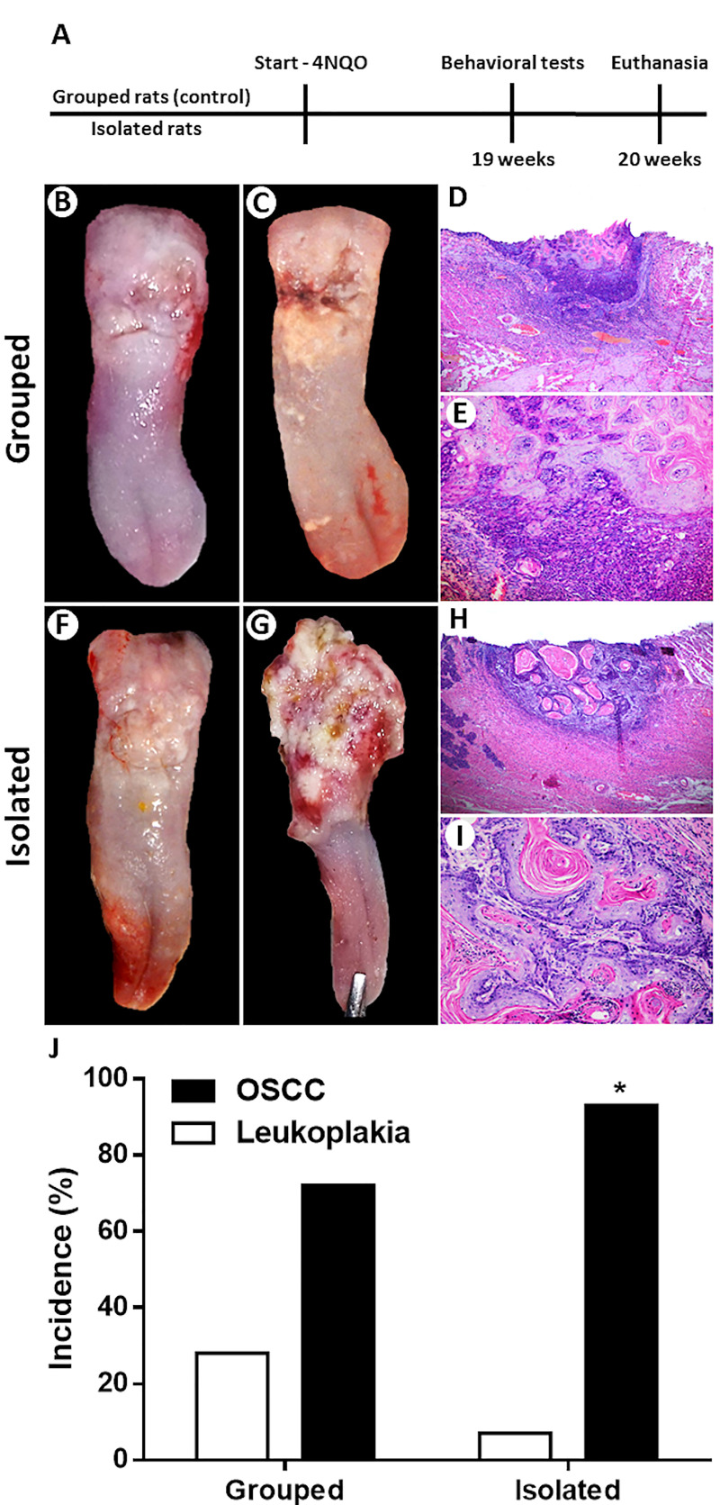
Experimental design (A). Clinicopathological characteristics of the OSCCs from grouped rats (B-E). White plates with a small ulcer (B). Yellowish white plaques with an infiltrative ulcer (C). Small-size OSCC (H&E; 50x) (D). Tumor epithelial cells surrounded by a chronic inflammatory infiltrate (H&E; 200x) (E). Clinicopathological features of the OSCCs from isolated rats (F-I). Irregular yellowish white plates (F). Extensive ulcer with reddish-white areas (G). Large-size OSCC (H&E; 50x) (H). Well-differentiated cells arranged in islands of varying size containing keratin pearls (H&E; 200x) (I). Social isolation stress increased the occurrence of chemically induced OSCC (J). Chi-square test showed that isolated rats had higher occurrence of OSCC than control rats. *p<0.05. Bar graphs represent the incidence rate of leukoplakia and OSCC for both groups.

