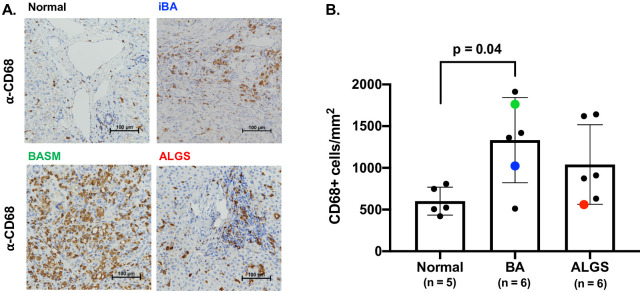
Fig 1. Increased hepatic macrophages in cholestatic liver disease.
Representative immunohistochemistry staining with the macrophage marker anti-CD68 in samples taken at the time of liver transplantation from the iBA, BASM, and ALGS patients also used for scRNA-seq are shown compared to a normal donor liver sample (A). Quantitative analysis of entire sections from wedge biopsies showed a significantly increased number of CD68+ macrophages in BA patients, with individual samples processed for scRNA-seq shown in blue (iBA), green (BASM) and red (ALGS) (B).

