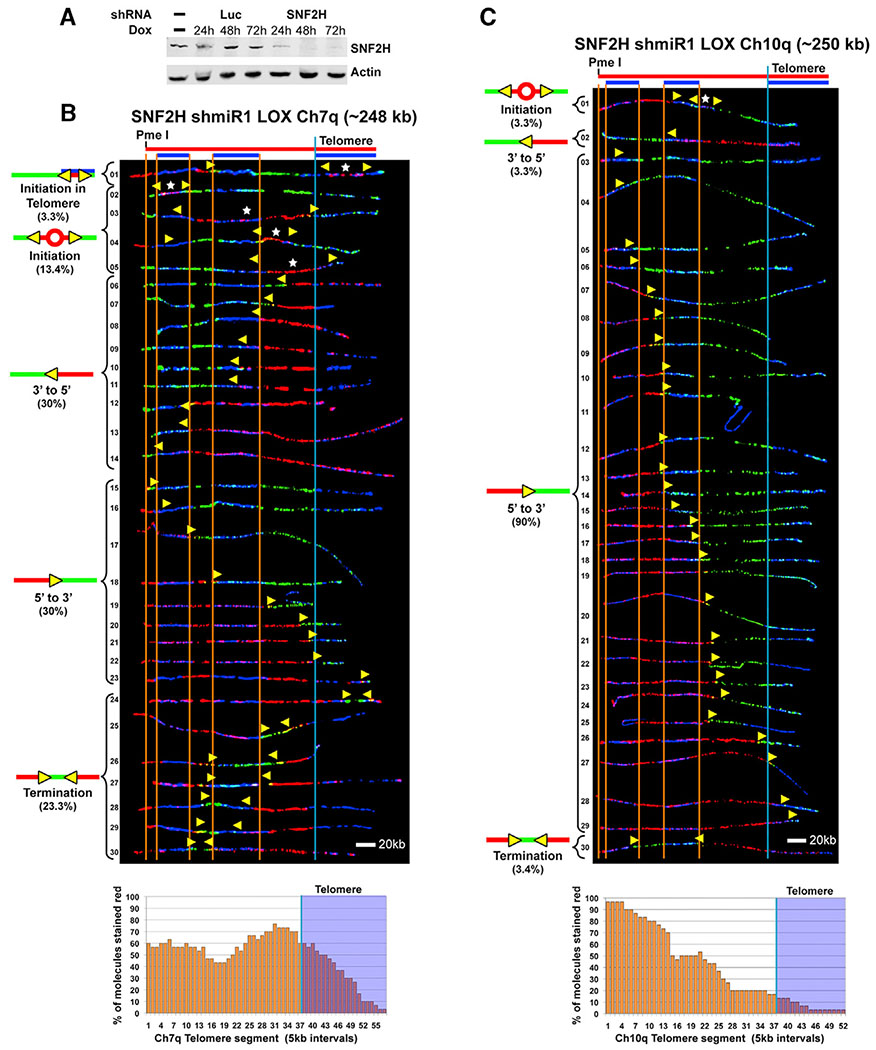
Figure 6. Knockdown of SNF2H Decreases Replication Initiation within Telomeres in LOX Cells.
(A) Inducible knockdown of SNF2H in LOX cells. Immunoblot of cell lysates of LOX cell lines stably expressing Dox-inducible SNF2H shRNA or Luciferase (Luc) control shRNA, showing a time course of induction over three days.
(B) SMARD analysis of the Ch7q telomere segment from LOX cells inducibly expressing shRNA against SNF2H. Cells were grown in the presence of 1 μg/mL Dox for 48 h prior to pulse-labeling with IdU, followed by labeling with CldU in the absence of Dox. Alignments of replicated molecules fully labeled with IdU (red) and CldU (green) are shown, collected from four independent samples stretched on slides (157 fully red- and 144 fully green-labeled molecules were also collected).
(C) SMARD analysis of the Ch10q telomere segment from LOX cells inducibly expressing shRNA against SNF2H as in (B). Alignments of replicated molecules fully labeled with IdU (red) and CldU (green) are shown, collected from two independent samples stretched on slides (55 fully red- and 58 fully green-labeled molecules were also collected). Vertical lines (orange and blue) demarcate the boundaries where FISH probes bind, as described in Figure 1. Symbols are as in Figure 1. Replication profile histograms are shown under the molecule alignments.

