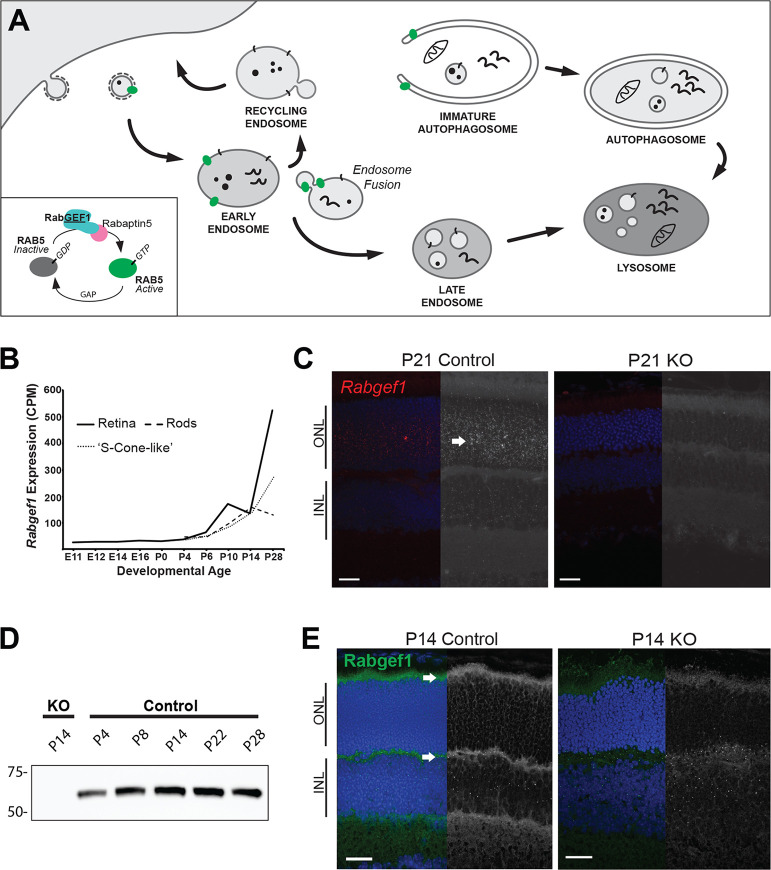
Fig 1. Spatiotemporal expression of Rabgef1 in the mouse retina.
(A) A schematic showing conserved functions of Rab5/RabGEF1 in endocytosis and autophagosome closure. (B) Rabgef1 expression in developing whole retina or flow-sorted rod and S-cone-like photoreceptors (identified from RNA-seq data in [30,36]). Nrl-GFP mice enabled purification of rod photoreceptors (Rods), whereas Nrl-GFP mice crossed with the cone-only Nrl-/- mice allowed sorting of S-cone-like photoreceptors [63]. (C) In situ hybridization profile of postnatal (P)21 control and Rabgef1-/- (KO) retina. Red/white punctate dots represent single Rabgef1 mRNA molecules. Arrow indicates high Rabgef1 transcripts in photoreceptor layer. Scale bar = 20 μm. ONL, outer nuclear layer; INL, inner nuclear layer. (D) Immunoblot analysis of P4 –P28 control and Rabgef1-/- P14 retinal lysates probed with anti-RabGEF1 antibody. The total protein loading control is included in S1 Fig. (E) Immunohistochemistry of P14 control and Rabgef1-/- retinal sections using anti-RabGEF1 antibody. Arrows indicate high RabGEF1 expression in photoreceptor inner segments and synaptic terminals. Only non-specific background staining (punctate dots), also observed in control, is detected in Rabgef1-/- retinal sections. DAPI was used for visualizing nuclei. Scale bar = 20 μm.

