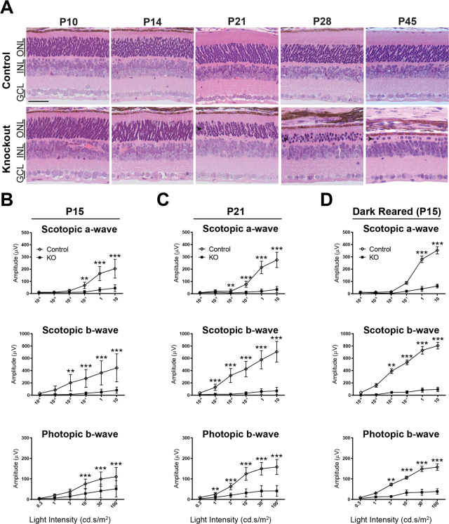
Fig 2. Rapid photoreceptor degeneration and loss of visual function in Rabgef1-/- mice.
(A) H&E staining of developing control and Rabgef1-/- retina, showing near complete ablation of photoreceptors (ONL) by postnatal day 45. Scale bar = 50 μm. ONL, outer nuclear layer; INL, inner nuclear layer; GCL, ganglion cell layer. (B, C) ERG stimulus intensity-amplitude functions in the control and KO mice reared in a 12h light/12h dark cycle and measured at postnatal day 15 and 21 (P15, P21), respectively. (D) ERG stimulus intensity-amplitude functions in control and Rabgef1-/- mice born and raised in the dark. Dark-reared Rabgef1-/- mice have similar ERG stimulus intensity-amplitude functions as animals reared in cyclic light. Asterisks indicate p-value < 0.05 (*), <0.01 (**) and <0.001 (***) as determined by a t-test using Prizm software. In panels B and C, n = 7–10 per genotype and in D, 2–3 per genotype. Error bars indicate SD.

