
Fig. 4: Additional histologic changes in AGMs infected with SARS-CoV-2.
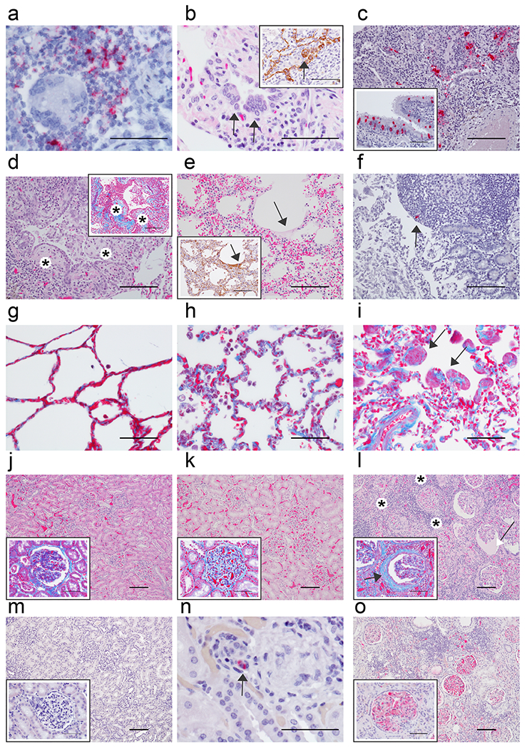
Pulmonary lesions in representative AGMs 5 dpi (a-e) and immunohistochemistry (IHC) of the (f) duodenum. a) Genomic SARS-CoV-2 RNA (red) detected by in situ hybridization in mononuclear cells near a multinucleated giant cell associated with acute bronchiolitis, 60x. b) Syncytial cells (arrows) within a terminal bronchiole, 60x and inset with pan-cytokeratin IHC positive syncytial cell indicating epithelial origin (arrow) within terminal bronchiole, 60x. c) SARS-CoV-2 positive IHC labeling (red) associated with acute bronchiolitis, 20x and in the inset SARS-CoV-2 IHC positive respiratory epithelium of the bronchus, 40x. d) Terminal bronchioles with multiple luminal protrusions of loose collagen covered by respiratory epithelium that is reminiscent of early formation of bronchiolitis obliterans organizing pneumonia (BOOP)-like lesions (*), 20x and inset serial section of tissue stained with trichrome highlighting immature collagen (*) (blue) 20x. e) Loss of alveolar architecture, marked expansion of septa and formation of faint hyaline membranes (arrow), 20x and inset IHC positive pan cytokeratin (brown) of hyaline membranes from serial section of tissue (arrow), 20x. f) IHC SARS-CoV-2 positive (red) mononuclear cell within the peyer’s patches of the duodenum (arrow), 20x. g) Trichrome stain of AGM alveolar septate basement membrane (blue) from a SARS-CoV-2 naïve AGM, 40x. h) Trichrome stain of alveolar septate with collagenous expansion (blue) from a SARS-CoV-2 AGM 5 dpi, 40x. i) and AGM 57 dpi and focal smooth muscle hyperplasia (arrows), 40x. j-o) Comparison of kidneys from naive AGMs, AGMs 5 dpi, and 57 dpi SARS-CoV-2. j) Renal congestion with NSL in naïve SARS-CoV-2 AGM, 10x and higher magnification in the inset of glomerulus with trichrome stain with no significant findings (NSF), 40x. k) Renal congestion with NSF in AGM 5dpi, 10x and higher magnification in the inset of glomerulus with trichrome stain with mild glomerular fibrosis, 40x. l) Marked renal interstitial lymphocytic inflammation (*) with glomerulopathy and expanded bowman’s space (white arrow) in AGM 57 dpi, 10x and higher magnification in the inset of glomerulus with trichrome stain with interstitial inflammation (*), glomerular fibrosis (blue), and marked periglomerular fibrosis (black arrow), 40x. m) Fibrin IHC negative AGM kidney 10x and higher magnification of fibrin negative glomerulus, 40x. n) IHC SARS-CoV-2 positive (red) mononuclear cell (arrow) within renal interstium of AGM 5 dpi, 60x. o) Fibrin IHC positive (red) multifocal within glomerular capillaries and renal interstitium of AGM 57 dpi, 10x and high magnification of fibrin positive glomerulus, 40x. H&E staining (b, d, e, j, k, & l), IHC labeling for anti-SARS-CoV-2 antigen (red) (c, c inset, f, & n), IHC labeling for anti-fibrin antigen (red) (m, m inset, o & o inset), IHC labeling for anti-pan cytokeratin (brown) (b inset and e inset), SARS-CoV-2 in situ hybridization (a), Trichrome (d inset, g, h, i, j inset, k inset, & l inset). Images captured at 10x (j, k, l, m & o), 20x (c, d, d inset, e, e inset & f), 40x (c inset, g, h, i, j inset, k inset, l inset, m inset & o inset), 60x (a, b, b inset, & n). Scale bar 100um (c, d, d inset, e, e inset, f, j, k, l, m & o) 50um (a, b, b inset, c inset, g, h, i, j inset, k inset, l inset, m inset, n, & o inset).