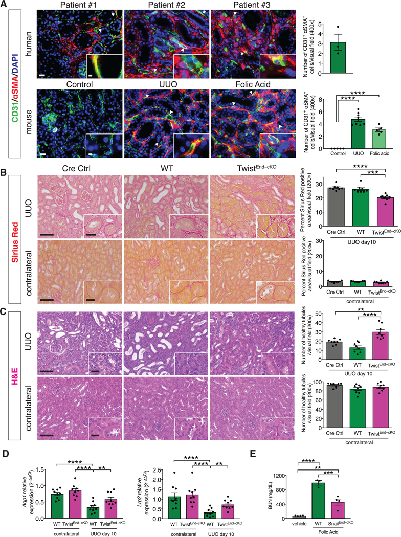
Figure 1. Conditional deletion of Twist1 in endothelial cells ameliorates UUO-induced kidney fibrosis.

(A) Immunolabeling for CD31 and αSMA in kidneys from human biopsies of obstructive and allograft nephropathies, n = 3 patients (top), and from UUO and Folic acid-induced experimental models of kidney fibrosis, n = 8 and n = 5 mice for each group, respectively (bottom). Scale bars: 20 μm. Insets: 5 μm. DAPI: nuclei. (B) Representative images of Sirius Red staining and quantification of Sirius Red positive area in UUO (top) and contralateral (bottom) kidneys from the indicated experimental groups. Cre Ctrl n = 8 mice, WT n = 8 mice, TwistEnd–cKO n = 8 mice. (C) Representative H&E images and quantification of the number of healthy tubules in UUO (top) and contralateral (bottom) kidneys from the indicated experimental groups. Cre Ctrl n = 8 mice, WT n = 8 mice, TwistEnd–cKO n = 8 mice. Scale bars: 100 μm. Insets: 25 μm. (D) Relative transcript levels of Aqp1 (which encodes Aquaporin 1) and Lrp2 (which encodes Megalin) in the kidneys of the indicated experimental groups. WT and TwistEnd–cKO n = 9 mice for each group. (E) Blood urea nitrogen (BUN) levels. Vehicle n = 4 mice, WT n = 3 mice, SnailEnd–cKO n = 4 mice. Data are presented as mean ± s.e.m. One-way analysis of variance (ANOVA) with Dunnett’s post-hoc analysis (A) or Tukey post-hoc analysis (B–E). **P < 0.01, ***P < 0.001, ****P < 0.0001.
