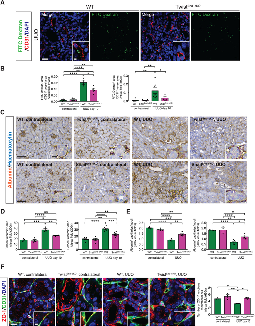
Figure 4. Blocking EndMT limits vascular leakage.

(A) Immunolabeling for CD31 and visualization of FITC-conjugated dextran in kidneys from the indicated experimental groups. Scale bars: 20 μm. DAPI: nuclei. (B) Quantification of the ratio of FITC-dextran+ area per CD31+ vessel area in UUO and contralateral kidneys of the indicated experimental groups. WT n = 3 mice, TwistEnd–cKO n = 4 mice; WT n = 7 mice and SnailEnd-cKO n = 5 mice. (C–D) Immunohistochemistry analysis of albumin in kidneys from the indicated experimental groups (C) and respective quantification (D). WT n = 3 mice, TwistEnd–cKO n = 4 mice; WT n = 7 and SnailEnd-cKO n = 5 mice. Scale bars: 100 μm. Insets: 25 μm. (E) Quantification of peritubular patented capillary density performed on the albumin immunohistochemistry analysis presented in (C). WT n = 3 mice, TwistEnd–cKO n = 4 mice; WT n = 5 and SnailEnd-cKO n = 3 mice. (F) Maximum intensity orthogonal projections of ZO-1 and CD31 co-immunolabeling and respective quantification of kidneys from the indicated experimental groups. All groups, n = 3 mice for each group. Scale bars: 30 μm. Insets: 7.5 μm. Data are presented as mean ± s.e.m. One-way analysis of variance (ANOVA) with Tukey post-hoc analysis. *P < 0.05, **P < 0.01, ***P < 0.001, ****P < 0.0001.
