
Figure 6. RtcR Oligomerizes on Binding tRNA Fragments Ending in Cyclic Phosphate.
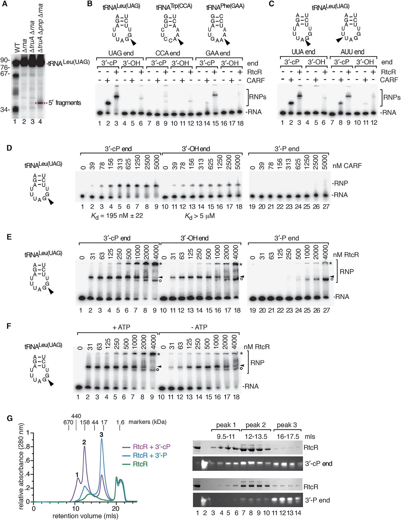
(A) RNA from the indicated strains was subjected to northern blotting to detect tRNALeu(UAG) 5′ halves.
(B) 32P-labeled 5′ tRNA halves (1 nM) ending after the anticodon in 2′, 3′-cyclic phosphate (lanes 1–3, 7–9, and 13–15) or 3′-OH (lanes 4–6, 10–12, and 16–18) were incubated with no protein, 0.5 μM CARF domain, or 0.5 μM RtcR. Reactions contained 1 mM ATP. RNA-protein complexes (RNPs) were separated from naked RNA in native gels.
(C) Similar to (B), except that 5′ tRNALeu(UAG) halves ended at the indicated positions.
(D and E) 5′ tRNALeu(UAG) halves ending after the G of the anticodon were incubated with the indicated concentrations of CARF domain (D) or RtcR (E). RNAs ended in 2′, 3′-cyclic phosphate (lanes 1–9), 3′-OH (lines 10–18), or 3′-phosphate (lanes 19–27). In (E), arrowheads denote the first complexes formed, while asterisks denote complexes that could represent oligomers. Circles, complexes that form on all three RNAs at the highest RtcR concentrations. In (D) and (E), the samples were fractionated in two gels. Binding reactions in (E) contained 1 mM ATP.
(F) tRNALeu(UAG) halves ending in cyclic phosphate were mixed with the indicated concentrations of RtcR with or without 1 mM ATP. Complexes are designated with arrowheads, asterisks, and circles as in (E).
(G) Size exclusion chromatography was performed on RtcR (48 μM) alone or bound to 48 μM 5′ tRNALeu(UAG) halves ending in 3′-phosphate or 2′, 3′-cyclic phosphate. Left, overlay of chromatograms. Right, proteins and RNA in the indicated peaks were fractionated in SDS-PAGE and denaturing polyacrylamide gels, respectively. The complex eluting at 20 mL is ATP from the binding reaction.
See also Figure S6.