
Figure 4.
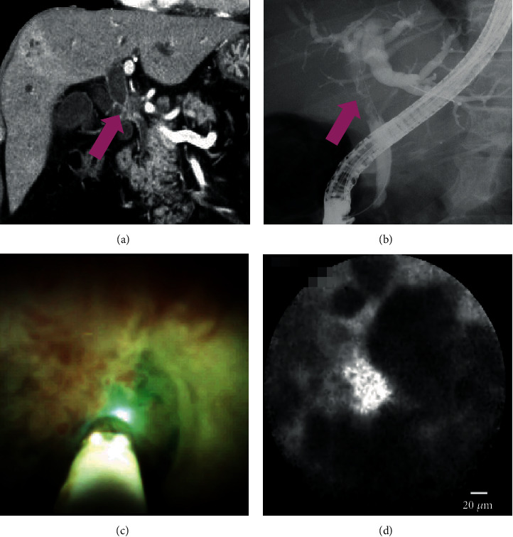
A case of malignancy: (a, b) computed tomography and cholangiography showing the biliary stricture in the hilar bile duct (pink arrow); (c) cholangioscopy showing an irregular papillogranular surface; (d) pCLE under the direct view of POCS showing dark glandular structures with irregular margins, suggestive of malignancy. Finally, the histological examination demonstrates adenocarcinoma.