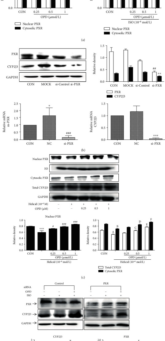
Figure 11.

The expression of CYP2J3 decreased after the PXR was knocked down. (a) OPD induces PXR nuclear translocation in H9c2 cells. ∗P < 0.05, ∗∗P < 0.01 compared with the control group. (b) The expression of CYP2J3 decreased after the PXR was knocked down, ∗∗∗P < 0.001, ###P < 0.001 versus the NC group. The effect of OPD on the transcription of the genes encoding CYP2J3 in HF H9c2 cells transfected with siPXR. aP < 0.01, dP < 0.01 versus the si-control group. bP < 0.05, cP < 0.05 versus the si-control group. eP < 0.001, fP < 0.001 versus the si-control group. (c) The expression of CYP2J3 decreased after the PXR was knocked down.∗∗∗P < 0.001 versus the control group, #P < 0.05, ###P < 0.001 versus the Helicid group. aP<0.05, bP<0.01 versus the control group. cP<0.05, dP<0.05, eP<0.05, fP < 0.05 compared with the Helicid group. (d) The effect of OPD on the expression of CYP2J3 and PXR after PXR was knocked down in HF H9c2 cells. aP < 0.01, dP < 0.01 versus the si-control group. bP < 0.05, cP < 0.05 versus the si-control group. eP < 0.001, fP < 0.001 versus the si-control group, n = 3 per group.
