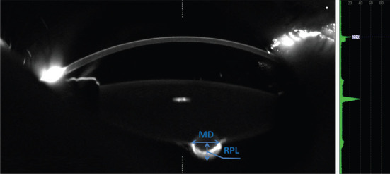
Figure 1. Scheimpflug image of the horizontal cross-section of the lens.

MDL was defined as the peak distance between the margins of both sides of the posterior lesion in all Scheimpflug image. The RPL was defined as the perpendicular distance from the center of the lens posterior surface to the focal protrusion apex.
