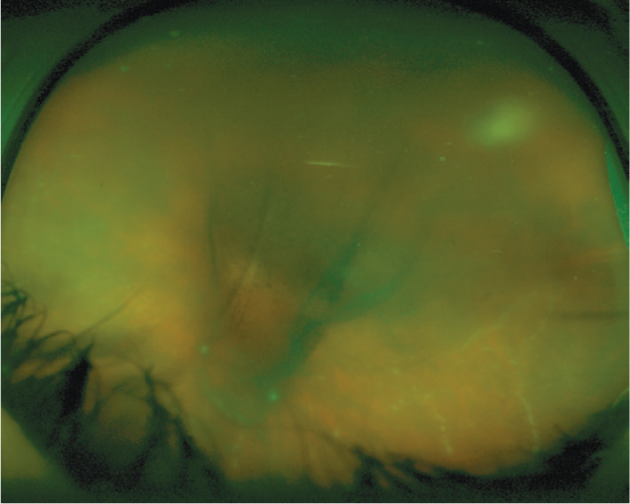
Figure 1. Wide-angle fundus photograph of the left eye (patient 1) showing dense central vitreous opacities obscuring the optic disc and the posterior pole with string-of-pearl vitreous opacities in inferior temporal part of the vitreous cavity.

A fuzzy white retinal lesion is also noted in superotemporal periphery.
