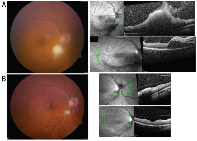
Figure 3. Color fundus photograph of the right eye (patient 3) at presentation (A) shows fluffy white retinal lesion along the inferotemporal arcade with preretinal extension and hazy view because of associated vitritis. Spectral-domain optical coherence tomography shows a hyperreflective dense retinal elevated lesion in the area of the white retinal infiltrate with vitreous infiltration and CME. Color fundus photograph of the right eye 10d later shows clear vitreous with marked regression of the white retinal lesion along the inferotemporal arcade (B). Spectral-domain optical coherence tomography shows resolution of the CME with fine hard exudates in the outer retinal layers and resolving retinal infiltrate.

