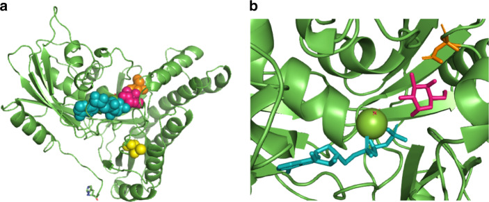
Fig. 2. The location of the two new disease-associated point variants in the human GALK1 structure.
(a) The three-dimensional structure of human GALK1 is shown in green. Adenosine triphosphate (ATP) (cyan) and galactose (hot pink) are shown bound in the active site. The two affected residues are shown: Asp46 (orange) and Thr352 (yellow). (b) A close-up of the active site showing how Asp46 binds, and helps orientate, the galactose molecule through C3-OH and C4-OH. The structure is based on PDB: 1WUU with gaps filled, selenomethionines converted to methionines, and AMP; PNP to ATP.5,40 Images created with PyMol.

