Fig. 1.
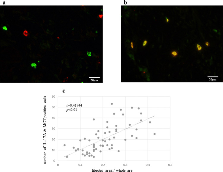
a IL-17A positive cells are red and CD4-positive cells are green. There were no double-positive cells (IL-17A and CD4) in the peritoneal disseminated lesion (original magnification ×400). b Double positive (IL-17A and MCT) cells are orange. Mast cells highly expressed IL-17A in the peritoneal dissemination (original magnification ×400). c The number of double (IL-17A and MCT)-positive cells correlated with the degree of fibrosis in peritoneal dissemination. p values were determined by Spearman’s correlation coefficient

