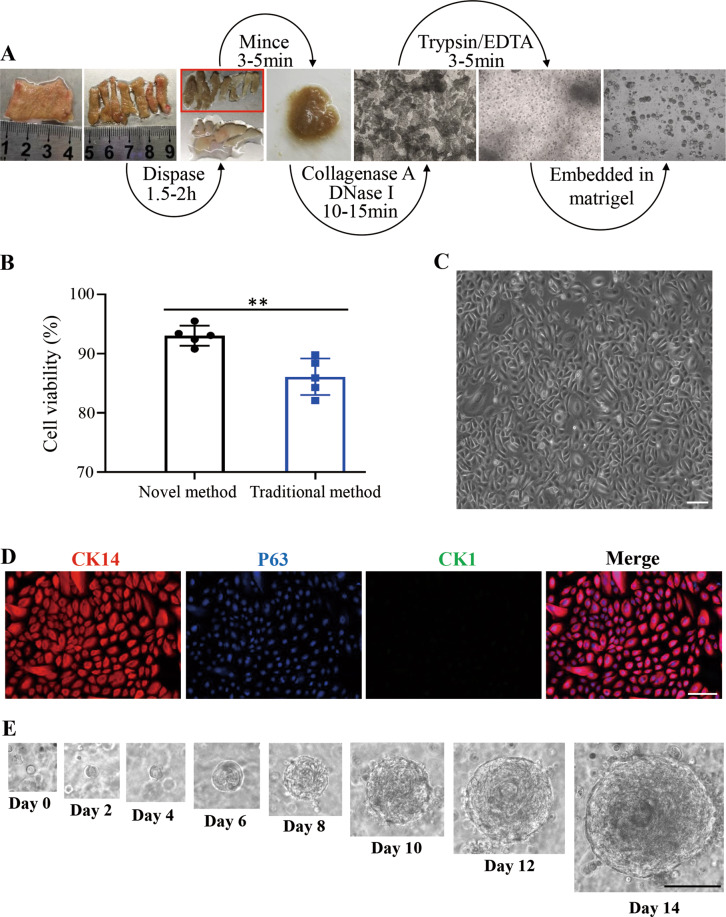
Fig. 1. A novel human epidermal cells isolation system and growth of hPEOs.
A Schematic representing isolation of epidermal cells from human foreskin tissue. B Comparison of the viability of cells derived from the traditional method and the novel method. Results are the mean ± SD from five independent repeated experiments. n.s., not significant (p > 0.05), *p < 0.05, **p < 0.01, ***p < 0.001. C Morphology of isolated human epidermal cells after attachment in a low-calcium, serum-free medium (EpiLife). D Immunofluorescence analysis of epidermal progenitor cell markers, CK14 and P63, in the attached epidermal cells. E Representative serial images of hPEOs growing at the indicated time points in the NaNBEFNoRWAFs medium. SD, standard deviation. Scale bar: 50 µm (C, D), 100 µm (E).

