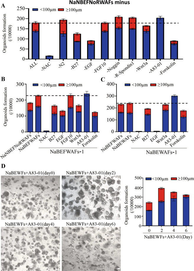
Fig. 2. Optimization of hPEOs cultures.
A The numbers of hPEOs after removal of individual factors from the NaNBEFNoRWAFs pool. B, C The numbers of hPEOs were counted after the removal of individual factors from the pool of NaBEFWAFs (B) and NaBEWAFs (C). D Representative images of hPEOs generated by various timings of A83-01 treatment in NaBEWFs medium. The hPEOs were counted on day 10. Results are the mean ± SD from three replicates from three independent repeated screening experiments. All screening experiments were performed with keratinocytes from three different donors. Scale bar: 100 µm (D).

