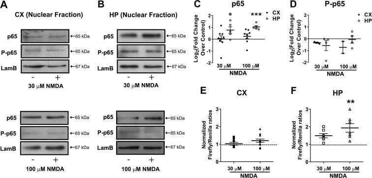
Fig. 1. NF-κB is activated in hippocampal, but not in cortical cultures after incubation with NMDA.
A, B Neuronal cultures were stimulated with 30 µM or 100 µM NMDA for 1 h and nuclear fractions were separated subsequently. Representative western blots of cortical (A) and hippocampal (B) culture-derived nuclear fractions after stimulation with 30 µM (top) or 100 µM (bottom). For each western blot, equal quantities of proteins were loaded and Lamin B1 (LamB) was used as a loading control. C, D Densitometric quantification of relative changes of p65 (C) and phospho-p65 (D) in the nuclear content, comparing stimulated (NMDA) vs. control (non-stimulated) condition in the same western blot. Calculated results obtained from 6 independent experiments (n = 6). Statistical significance was assessed by a two-tailed t-test (*p < 0.05; **p < 0.01). E, F Showing relative luciferase activity in cortical (E) and hippocampal (F) neurons after stimulation with 30 µM or 100 µM NMDA for 1 h (n = 6). Statistical significance was assessed by one-way ANOVA followed by Bonferroni post-test (**p < 0.01).

