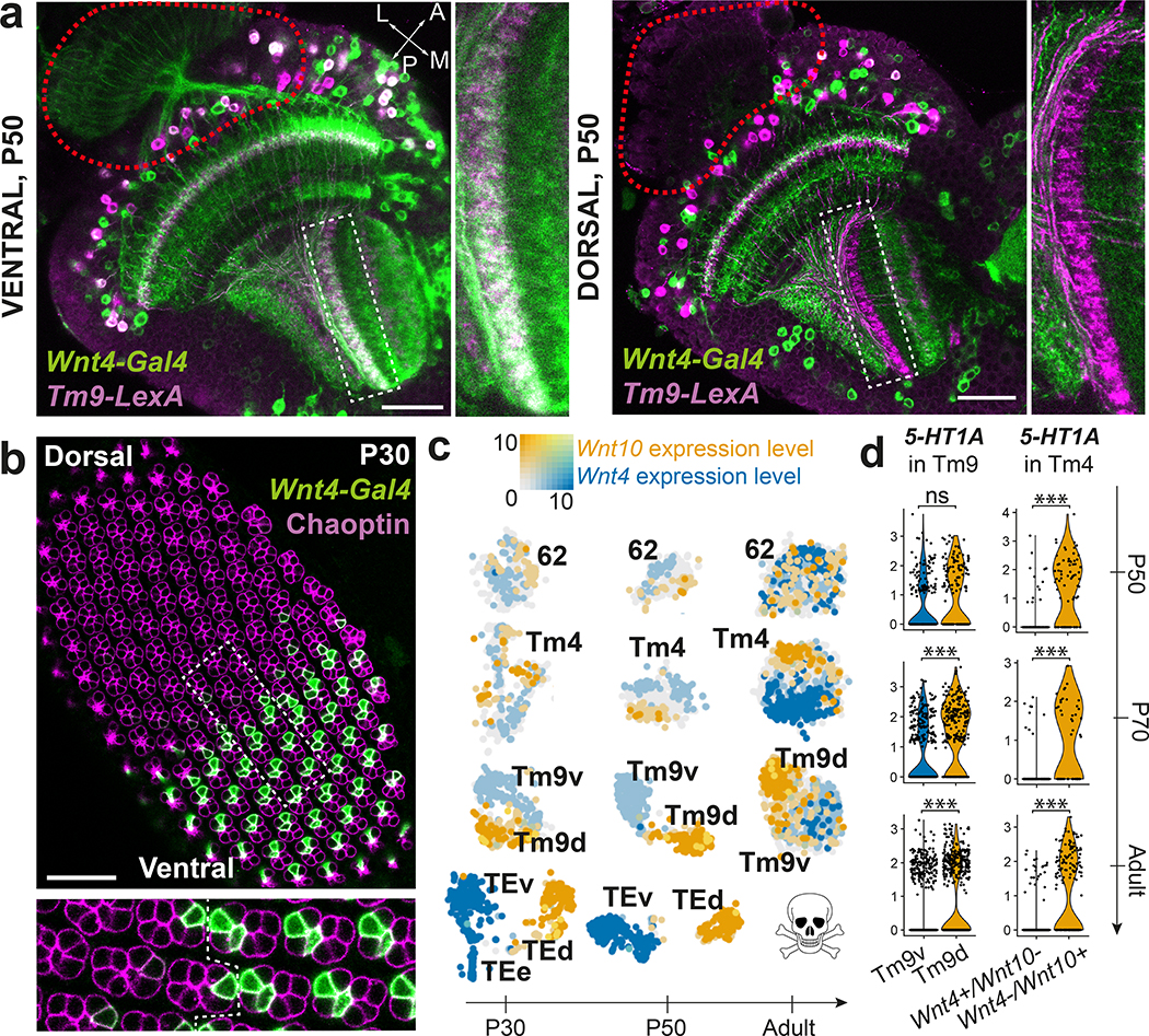
Figure 5: Dorsal and ventral visual circuits are partitioned by differential Wnt signaling.
a, Pattern of Wnt4-Gal4 and Tm9-LexA co-expression (white) at P50 in the ventral and dorsal part of the same optic lobe (n=8 brains). Red dashed line: location of photoreceptors. b, Wnt4-Gal4 expression pattern with anti-Chaoptin staining to mark photoreceptors in a P30 retina (n=6 eye discs). Dashed rectangle: inset. Dashed line within inset: equator of the retina. c, tSNE of the indicated clusters throughout development, with Wnt4 and Wnt10 log-normalized non-integrated expression levels. d, 5-HT1A differential expression between either Wnt4+/Wnt10- and Wnt4-/Wnt10+ Tm4 cells, or Tm9v and Tm9d cells (Methods). ***: adjusted p-value < 0.001 (P70 Tm9: 6×10⁻⁹, Adult Tm9: 3×10⁻¹⁸, P50 Tm4: 1×10⁻⁹, P70 Tm4: 2×10⁻⁷, Adult Tm4: 1×10⁻³⁴), ns: not significant, two-sided Wilcoxon Rank Sum test. Scale bars = 30 μm.

