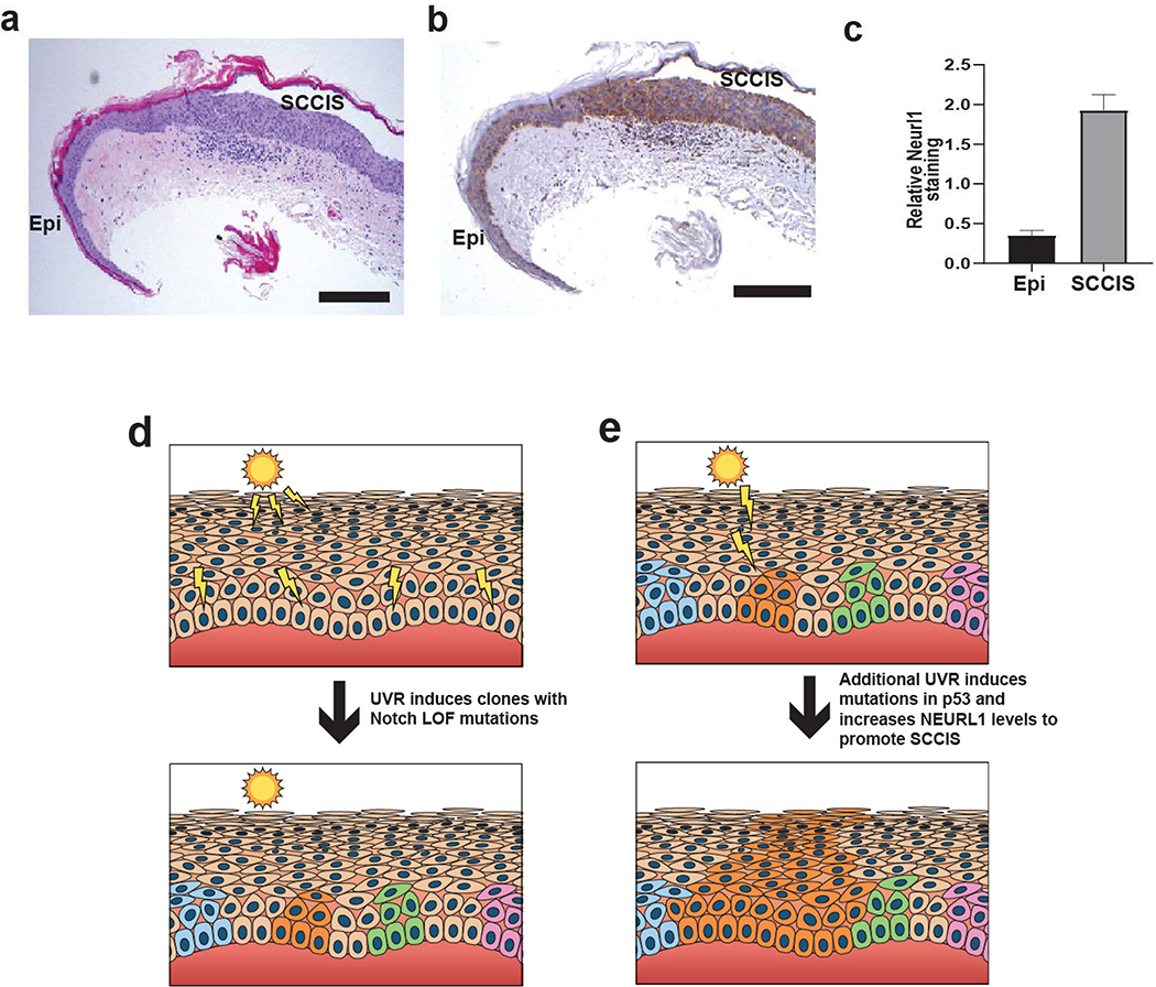
Figure 5. Immunohistochemical analysis of Neurl1 in epidermis and SCCIS with model of UV-induced SCCIS formation.
(a) H+E staining of representative specimen containing epidermis (Epi) and SCCIS. Scale bar = 0.25 mm. (b) Same representative immunohistochemical staining for Neurl1 showing higher levels of staining in SCCIS than in Epi. Scale bar = 0.25 mm. (c) Analysis of Nerul1b staining intensity. The relative staining intensity for Neurl1 is higher in SCCIS than adjacent epidermis. N = 10, p < 0.0001. (d) Scattered epidermal keratinocytes exposed to UVR acquire independent Notch LOF mutations and form small clones, clones highlighted in color. (e) A random clone acquires additional UV-induced mutations in p53 and/or increased levels of NEURL1 ubiquitin ligase that promotes progression to SCCIS manifested by full-thickness epidermal growth of lesional cells orange)

