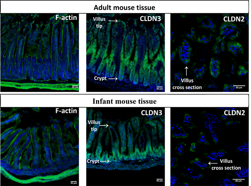
Fig. 3. – Infant and adult intestinal mouse tissue differ in morphology and tight junction localization.
In each representative immunofluorescence image, nuclei are stained with DAPI (blue) and the protein of interest appears in green. Cytoskeleton (F-actin) staining showed taller, more mature villi in adult tissue. While the barrier-forming Claudin 3 (CLDN3) appears throughout adult villi, it is localized exclusively to the crypts in infant tissue. Cross sections of villi show localization of Claudin 2 (CLDN2) localized differently in adult tissue (towards cellular junctions) compared to infant tissue (intracellularly).

