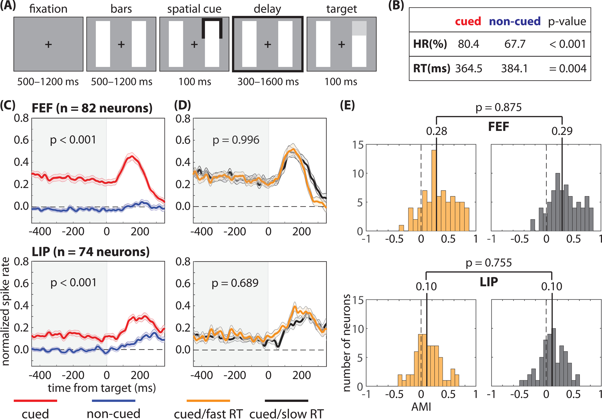
Figure 1. Pre-target differences in spike rates are not associated with differences in response times.

We recorded from the frontal eye fields (FEF) and the lateral intraparietal (LIP) region while monkeys completed (A) a spatial-cueing task. The animals demonstrated (B) both a higher hit rate (HR) and faster response times (RTs) when low-contrast targets occurred at the cued location relative to a non-cued location. (C) Pre-target spike rates (i.e., during the cue-target delay), averaged across neurons, were higher when receptive fields (RFs) overlapped the cued location; however, (D) we observed no differences in pre-target spike rates, averaged across neurons, between fast- and slow-RT trials (i.e., when RFs overlapped the cued location). (E) We also observed no differences in the median attentional modulation index (AMI) between fast- and slow-RT trials. The shaded area around each line represents the standard error of the mean. See also Figures S1–S4.
