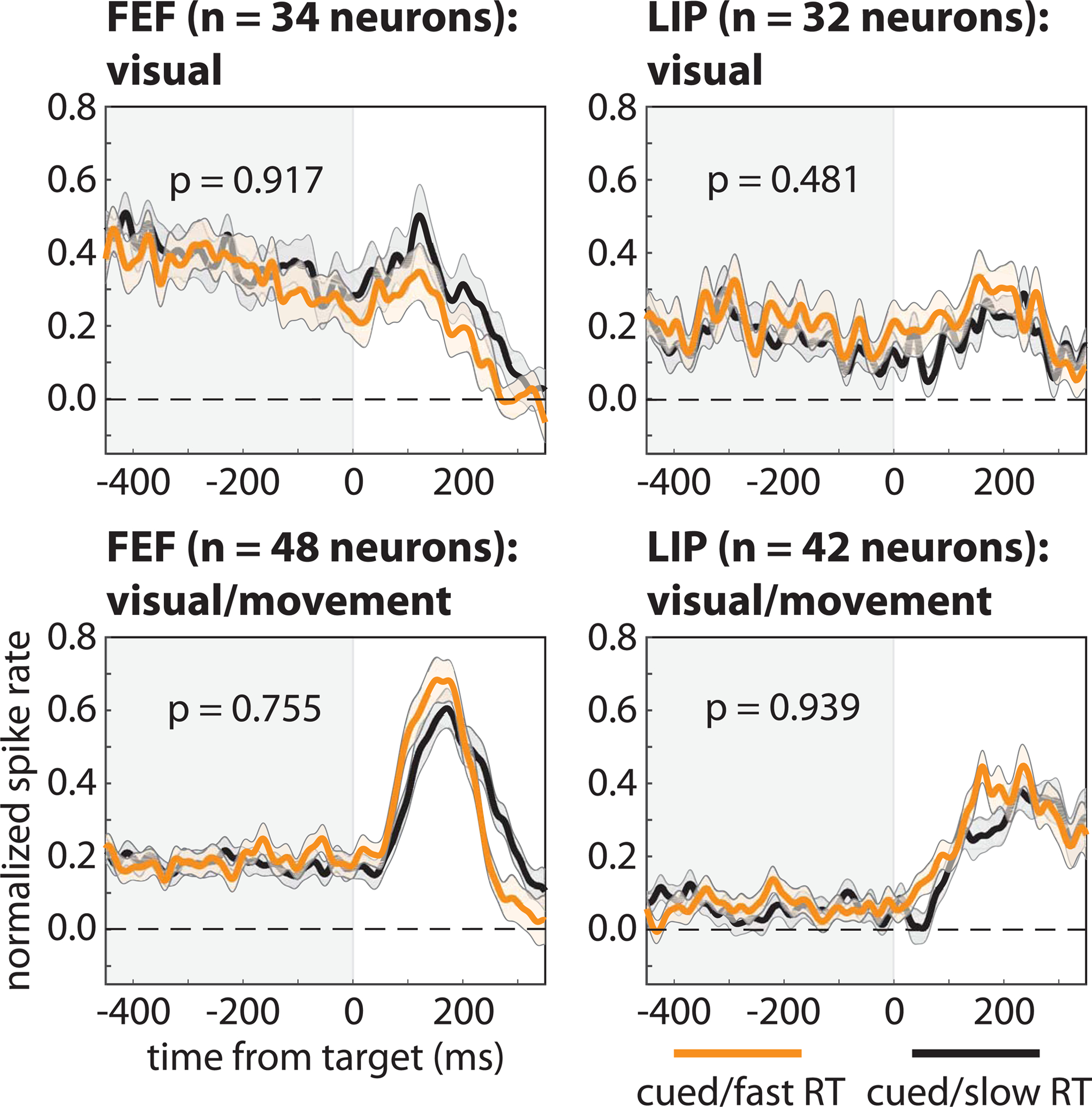
Figure 2. Pre-target differences in spike rates among functionally defined cell types are not associated with differences in response times.

We binned neurons in FEF and LIP into visual and visual-movement cell types, with the former only responding to visual stimulation and the latter both responding to visual stimulation and demonstrating saccade-related activity. Regardless of cell type and brain region, there we observed no differences in pre-target spike rates, averaged across neurons, between trials that resulted in either faster (orange lines) or slower (black lines) response times (i.e., when RFs overlapped the cued location). The shaded area around each line represents the standard error of the mean.
