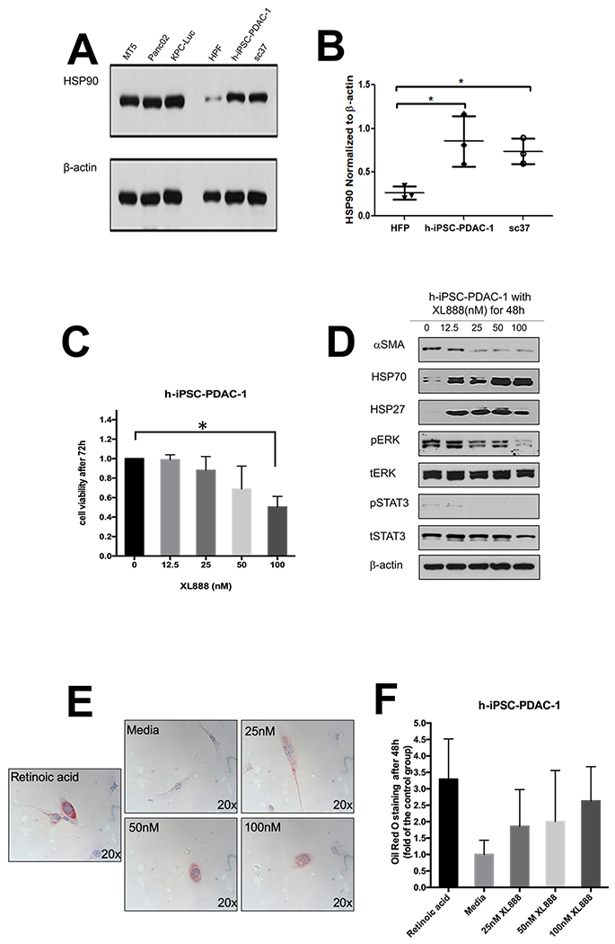
Figure 1. Hsp90 inhibition limits activation of PSC/CAF.
(A) Immunoblot analysis of Hsp90 expression in a panel of murine PDAC cell lines (MT5, Panc02, KPC-Luc), normal human pancreatic fibroblasts (HPF), immortalized PDAC-derived human PSC/CAF (h-iPSC-PDAC-1) and a primary PDAC patient-derived PSC/CAF culture (SC37). (B) Densitometry analysis from n=3 biological replicate blots. (C) MTT assay and (D) immunoblot of h-iPSC-PDAC-1 cells treated with increasing concentrations of XL888. (E) Oil Red O staining of h-iPSC-PDAC-1 cells following treatment for 48 hours with XL888. Al-trans retinoic acid treated cells served as a biologic positive control. For immunoblot analysis, β-actin served as a loading control. Error bars represent standard deviation of n=3 biologic replicates; *denotes p<0.05.

