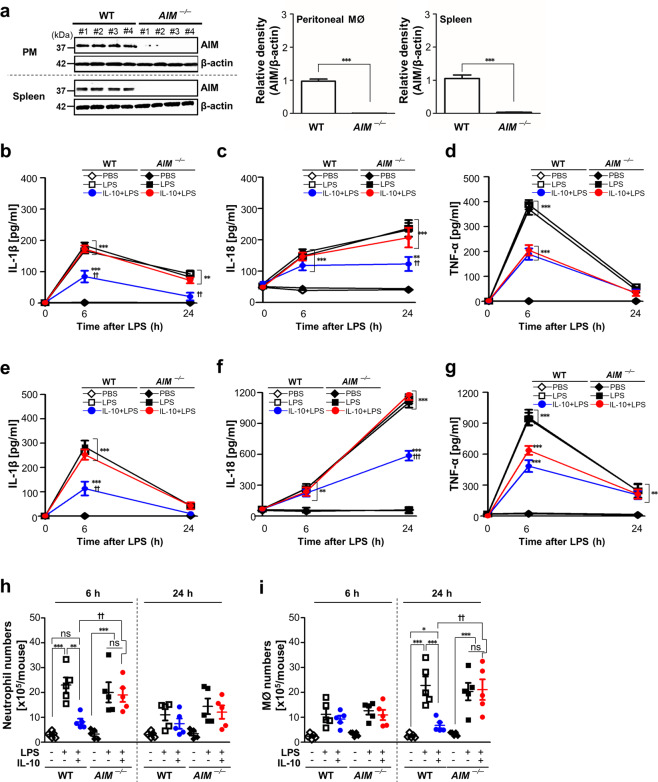
Fig. 7. In vivo inhibitory effects of IL-10 on IL-1β and IL-18 production and inflammatory cell recruitment in LPS-induced peritonitis were reversed in AIM−/− mice.
a Left: Immunoblot analysis of the indicated protein in lysates of peritoneal macrophages and spleen from wild type (WT) and AIM−/− mice (n = 4 mice). Right: The relative densitometric intensity was determined for each band and normalized to β-actin. b–i Where indicated, WT and AIM−/− mice were injected i.p. with 10 mg/kg LPS before administration of murine IL-10 (i.p., 30 μg/kg). Animals were euthanized at 6 or 24 h after LPS injection. ELISA was performed to quantify the abundance of IL-1β (b, e), IL-18 (c, f), and TNF-α (d, g) in peritoneal lavage fluid (PLF) and in serum, respectively. Numbers of neutrophils (h) and macrophages (i) in PLF were determined. Values represent the mean ± SEM of five mice per group. ns: not significant; **P < 0.01 and ***P < 0.001 compared with PBS control; ++P < 0.01 and +++P < 0.001 for AIM−/− mice versus WT mice treated with IL-10+LPS at a given time point.

