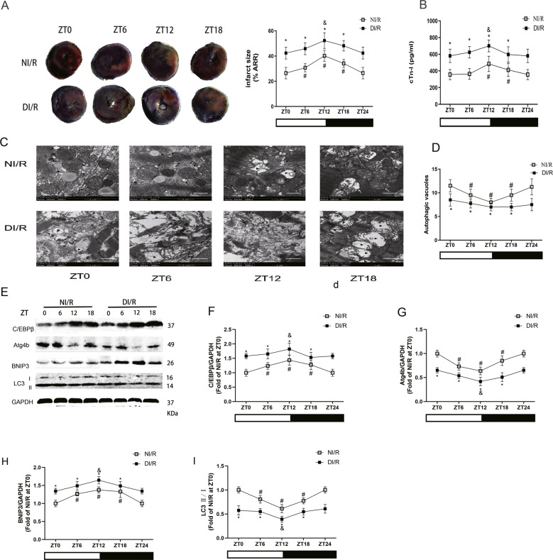
Fig. 2. The circadian dependence of MI/RI in diabetic rats was attenuated, which was associated with disordered rhythm of mitophagy.
A Infarct size was detected by TTC. Scale bar: 2 mm. B The serum level of cTn-I was detected by ELISA in non-diabetes or diabetes with I/R insult. C and D The ultrastructural changes and autophagic vacuoles of rat hearts were detected by TEM (MT: normal mitochondrion, white arrows: swollen mitochondrion, ▶: disorganized and vacuolated mitochondrion, black arrows: autophagosome/autophagic vacuole, Scale bar: 1 μm. E–I The protein levels of C/EBPβ (F), Atg4b (G), BNIP3 (H) and LC3 II/I (I) were detected by western blotting in the myocardial tissues of non-diabetes or diabetes with I/R insult. n = 6 per group. *P < 0.05 versus NI/R within ZT; #P < 0.05 versus ZT0 within NI/R; &P < 0.05 versus ZT0 within DI/R.

